Dry Eye Tests: Here is a list of all the available test for Dry Eye

Dry Eye Tests: 
Here is a list of all the available test for Dry Eye to date.

Patients with Dry Eyes initially may not think “a little dry eye” is a big deal. This is true for the majority of patients. However, if left untreated, many patients can progress to debilitating disease. In a small percentage of patients, though, dry eye can lead to true devastation and disability: having chronic pain with every blink can be very difficult to live with for some patients. 


Thus comprehensive testing is very important for any patient with dry eyes and their physician. Testing, though, for Dry Eye has been frustrating for patients and physicians alike. Many dry eye tests have a low sensitivity and specificity. 


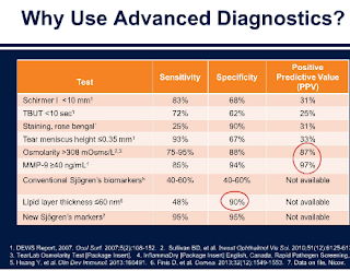
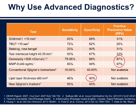
The most classic dry eye tests are noted here and their positive positive test that are true positives (Positive Predictive Value). As you can see the PPV of these classic tests are low.


1. Schirmer: 31%
2. Tear Break Up Time: 25%
3. Staining of Cornea: 31%
4. Tear Meniscus Height 33%


Thus new, innovative ways of diagnosing dry eye or meibomian gland dysfunction have been developed in recent years. 


The key issue in many of these tests is that they are expensive and not covered by insurances. Many patients cannot find some of the advanced tests in their doctors offices (especially as many of these tests are not reimbursed by insurances and thus a money loosing issue for offices.) Additionally offices often cannot even charge patients for these tests because of insurance regulations and thus many eyeMD offices are no longer doing many of these tests. 


Here is a comprehensive list of dry eye tests and the positives and negatives of each test. If you know of new ones or ones I overlooked, please let me know. 

Sandra Lora Cremers, MD, FACS


A. TEAR FILM STABILITY TESTS: A normal tear film should be continuously available from a combination of 3 key areas:
(1.) Aqueous from the Lacrimal Gland: this is decreased in patient with autoimmune diseases like Sjogren’s syndrome, Rheumatoid Arthritis, Lupus, etc.
(2.) Lipid/oil from the Meibomian Gland: this is decreased in patients with Meibomian Gland Dysfunction from Rosacea, chronic Blepharitis (inflammation of the eyelid margin from bacteria like Staphlococcus or mites like Demodex). 
(3.) Mucin from the Goblet Cells: decreased in patients who have had prolonged chlorine exposure, likely abused contact lenses for years, 

Blinking maintains the tear film continuity. However if you keep your eyes open long enough, without blinking, the tear film will start breaking up into dry spots we can see under the microscope. Your eye will feel uncomfortable forcing you to blink. In patients with dry eyes the tear film is unstable, and breaks up faster. Therefore the tear break up time in patients who have dry eyes is shorter.

1. Tear Film break-up time (TFBUT or FBUT)
Measures the interval between the individual’s last complete blink and the break-up of a patient’s tear film
  1. We use of a microscope called a slit-lamp, set on a bright light setting with a cobalt blue filter
  2. We Instill fluorescein into the lower fornix.
  3. Patient blinks several times and then stop.
  4. We measure the time between the last blink and the first appearance of a dark spot on the cornea (formation of a dry area) on the otherwise continuously stained tear film. 
  5. A tear break-up time of less than 10 seconds suggests a dry eye.

2. Non-invasive tear break up time (NIBUT): no Fluorescein is used & eye is not touched
Fluorescein in the tears may stimulate reflex tearing and may also result in changes to the tear film properties.

a. Instruments such as a Keratometer, hand-held Keratoscope or Tearscope are required to measure NIBUT.
b. A pre-rupture phase that precedes actual break up of the tear film can also be observed with some techniques. This pre-rupture phase is termed Tear Thinning Time (TTT). Measurement is achieved by observing the distortion (TTT) and/or break up (NIBUT) of a keratometer mire (the reflected image of keratometer grid). The clinician focuses and views the crisp mires, and then records the time taken for the mire image to distort (TTT) and/or break up (NIBUT).
c. NIBUT measurements are longer than fluorescein break up time. NIBUT values of less than 15 seconds are consistent with dry eyes.
d. Not very sensitive or specific.

B. TEAR VOLUME TESTS
Tear volume and production are components of tear film stability that can be affected by dry eye. One common method for determining tear volume is the Schirmer test. 

1. Schirmer test: there is actually a Schirmer I and Schirmer II test: this describes Schirmer II

  1. Instil a drop of local anaesthetic into the eye, (optional).
  2. Prepare a filter paper (5mm x 35mm with folded end).
  3. Gently dry the eye.
  4. Apply the filter paper with the folded end hooked onto the lower lid margin at the junction between the middle and outer third (take care not to touch the cornea).
  5. Tell patient to keep their eye open and blink normally.
  6. Measure the amount of wetting after 5 minutes: 13-15mm wetting rules out a dry eye; 6-10mm is borderline and less than 6mm indicates dry eye.
  1. The filter paper strips can cause reflex tearing and may require the use of anaesthetic agents: if not anesthetic agent is used, it measure reflex tearing & is called Schirmer’s I test. These test have fallen out of favor after published reports showed lack of correlation with true dry eye patients. 

2. Phenol Red Test
A cotton thread impregnated with phenol red dye is used. Phenol red is pH sensitive and changes from yellow to red when wetted by tears.

  1. The crimped end of a 70mm long thread is placed in the lower conjunctival fornix.
  1. After 15 seconds, the length of the colour change on the thread – indicating the length of the thread wetted by the tears -is measured in millimetres.
  1. Wetting lengths should normally be between 9mm and 20mm. Patients with dry eye have wetting values of less than 9mm.

3. Tear Prism Height Test
A significant amount of information about tear quantity can be gained simply from observing the heights of the upper and lower tear menisci with the slit-lamp biomicroscope.

There are two techniques used when measuring the height of tear prisms. 
a. The first measures the tear meniscus formed on the lower lid margins to give a useful guide to tear volume. This simple technique employs the slit lamp biomicroscope. Excessive or prolonged use of illumination should be avoided to prevent artificial drying of the tear prism.
b. The second technique is to compare the tear prism height with the illuminated slit width by setting the slit horizontally in alignment with the lower lid margin, altering the slit width until it appears to match the height of the tear prism. Heights of less than 0.2mm indicate reduced tear fluid quantity.
c. Observation of the meniscus profile is also extremely helpful. A regular tear meniscus is typically observed in a healthy eye while a meniscus with a scalloped edge is often associated with a dry eye.
d. This is done on most patients by most dry eye specialists. 


C. STAIN TESTS
Corneal and conjunctival staining serves as an indicator of the health of the ocular surface. 

1. Rose Bengal Test (No longer widely used)
End-organ damage to conjunctival and corneal epithelial cells can be assessed via Rose Bengal ocular surface staining, which identifies areas of devitalised tissue:

  1. Instill a drop of Rose Bengal dye into the inferior fornix of the unanesthetised eye.
  2. Ask the patient to blink twice, to spread the red stain over the conjunctiva and cornea.
  3. The ideal time to measure the presence of staining is approximately 3 to 5 minutes after instilling the drops.
  4. Score the staining using a slit-lamp: a pattern of exposure zone (interpalpebral) corneal and bulbar conjunctival staining is typically seen with aqueous tear deficiency.

2. Lissamine Green (less widely used)
Lissamine green staining like Rose Bengal, colours any desiccated and dying cells on the ocular surface. However, lissamine green may be better accepted by patients as it lacks the slight burning or stinging sensation typically found with rose Bengal.

3. Fluorescein (most commonly used)
Fluorescein staining penetrates areas of the corneal epithelium and conjunctival epithelium where intercellular junctions are disrupted.


D. HYPEROSMOLARITY TEST

An increase in solute particles (proteins) within the tear film, or Hyperosmolarity of the tear film, is recognized as an important pathogenetic factor in dry eye syndrome (DES). Tear hyperosmolarity is a key feature of “ocular surface dryness”. 

1. Tearlab Osmolarity System: a new user-friendly tool that only needs tiny volumes for analysis and determines hyperosmolarity semi-automatically.
a. The disposable probe, touched onto the lower tear meniscus at the lid margin, collects a nanolitre sample of tears, which is analysed within seconds to provide the clinician with an osmolarity reading. Normal values lie around 304mOsm/kg while values over 320mOsm/kg indicate dry eye. 
b. Most eyeMD offices are starting to stop using TearLab as they are loosing money on this product.
c. My office uses TearLab but possibly for only a short time more for this reason. 


2. InflammaDry by RPS: very exciting technology. 
InflammaDry® is the first and only, rapid, in-office test that detects MMP-9, an inflammatory marker that is consistently elevated in the tears of patients with dry eye disease.1 Using direct sampling microfiltration technology, InflammaDry® accurately identifies elevated levels of MMP-9 protein in tear fluid samples taken from the inside lining of the lower eyelid, the palpebral conjunctiva.
InflammaDry® is a disposable, low cost test, that requires no additional equipment to administer or interpret results. Using four simple steps, InflammaDry® test results are achieved in just 10 minutes, aiding in the diagnosis of dry eye before the patient leaves the office.
This issue, though, for many offices is again the issue of re-imbursement. Many eyeMDs are seeing that most insurances do not cover the cost of the test. 

E. OTHER

1. LipiView: exciting new technology but we still do not know what all the data it provides really means in terms of prognosis. It is expensive as well: range $1000-2000 depending if Lipiflow is performed. It available near our office


2. Goblet Cell Density: rarely done but helps determine amount of goblet cells of the conjunctiva which produce mucin.
a. Usually only used for patients in a particular study. We used Goblet Cell Density in a study on pterygium patients.

3. SJO: Sj
ögren’s Test: 
Sjögrens is an important diagnosis for eye doctors to NOT miss. All doctors should ask their patients if they have dry mouth and/or arthritis if the patient has any dry eye complaint (like foreign body sensation, rebound tearing, redness). 


Sjögrens is a serious diagnosis because of it association with many other issues, in particular, Non-Hodgkin lymphoma which can happen in 5% of people with the syndrome.

It’s estimated that people with Sjögren’s syndrome are 44 times more likely to develop non-Hodgkin lymphoma than people without Sjögren’s .
Non-Hodgkin lymphoma is a cancer of the lymphatic system. The lymphatic system is a series of vessels and glands (the lymph nodes) that are spread throughout your body, much like your blood vessels.
If you have Sjögren’s syndrome, you should be aware of the main early symptom of non-Hodgkin lymphoma, which is a painless swelling in a lymph node (gland) – usually in the neck, armpit or groin. Report any swollen lymph nodes to your MD.

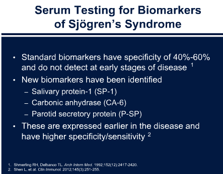
The Sjö test has a Higher Specificity and Sensitivity compared to 
40-60% sensitivity of anti-Ro or anti-La (older tests)
Sjo tests include: Salivary Protein-1 (SP-1); Carbonic Anhydrase (CA-6); Parotid Secretory Protein (P-SP)


More information below on Sjogrens:


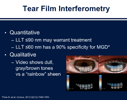
4. Lipid Analysis: Lipid Layer Thickness <60nm2 is a significant sign that the meibomian glands are not functioning as they should. 


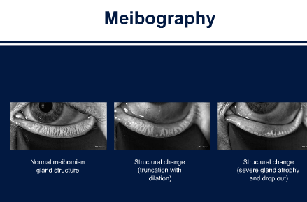
5. Meibography: Shows meibomian glands and their drop out.
It can convince patients they need treatment.
The machine to be able to show patients is still coming to be able to show patients. Harvard and many academic centers do not have this machine yet.

6. Optical Coherence Tomography on the Cornea is starting to be used more and more to assess tear film. 


7. Tear Lactoferrin: coming soon: new test

http://teardiagnostics.com/doctors/lactoferrin/
Lactoferrin (Lf), also known as lactotransferrin, is a multifunctional, iron binding glycoprotein that is widely present in mammalian tears, saliva, milk and mucosal secretions. In tears, Lf is one of the eye’s main immunological defense agents and plays a well-documented and critical role in the maintenance of ocular surface health.
Lactoferrin’s biological actions include:
  • Modulation of ocular inflammatory response
  • Regulation of normal cell growth
  • Protection against infections by inhibiting viral, bacterial or fungal growth
Secreted directly by the epithelial acinar cells of the lacrimal gland, lactoferrin is considered a bacteriostatic in tear film and is necessary for the formation of the principle natural ocular antibiotic, lysozyme.
As a biomarker, lactoferrin has been studied extensively and has long been established as a highly accurate test in assessing the secretory function of the Lacrimal Gland.
In the tear film, lactoferrin accounts for approximately 25% of total proteins by weight. Lower tear film lactoferrin levels are reported in dry eye, herpes simplex keratitis and systemic infections such as HIV and Hepatitis C.

———–
http://www.uptodate.com/contents/sjogrens-syndrome-beyond-the-basics

Patient information: Sjögren’s syndrome (Beyond the Basics)

SJÖGREN’S SYNDROME OVERVIEW
Sjögren’s syndrome is a chronic disease in which the body’s immune system mistakenly attacks glands that produce moisture in the eyes, the mouth, and elsewhere in the body. The most common symptoms of Sjögren’s syndrome are dry eyes and dry mouth. The symptoms in Sjögren’s syndrome are commonly divided into “benign” and “systemic” (or extraglandular). Although the symptoms are called “benign,” the complaints of dry or painful eyes/mouth, fatigue, and muscle ache are considered by many patients to be their greatest cause of disability.
This type of disease is called an autoimmune disease, meaning that the body’s immune system attacks its own tissues or organs. Sjögren’s syndrome can affect one or more organ systems including the skin, lung, heart, kidney, and nerves. Patients may have anemia, low white blood cell or platelet count, as well as increased risk of developing of lymphoma. Some people have Sjögren’s syndrome as well as another autoimmune condition, such as systemic lupus erythematosus, rheumatoid arthritis, or scleroderma.
Although there is no cure for Sjögren’s syndrome, a number of treatments are available. This article discusses the possible causes, signs, symptoms, diagnostic process, and treatments of Sjögren’s syndrome.
SJÖGREN’S SYNDROME CAUSES
Sjögren’s syndrome is thought to be caused by the body’s own immune system. Lymphocytes are a type of white blood cell in the body’s immune system that normally help to protect the body from infection. In Sjögren’s syndrome, these cells are thought to damage the glands that produce tears and saliva.
A person who develops Sjögren’s probably inherits the risk from one or both parents and is then exposed to some type of environmental trigger (eg, a viral infection), but the exact cause is not known (see “Pathogenesis of Sjögren’s syndrome”). Thus, both genetic factors and non-genetic factors play a role. Genome sequencing of Sjögren’s syndrome patients in both the US and Asia has added greatly to our understanding of the genetic factors by identifying at least five different risk-related major gene regions.
SJÖGREN’S SYNDROME SYMPTOMS
The classic symptoms of Sjögren’s syndrome are dry mouth (due to decreased production of saliva) and dry eyes (due to decreased production of tears). Symptoms of Sjögren’s syndrome can develop in otherwise healthy people, especially older adults. (See “Clinical manifestations of Sjögren’s syndrome: Exocrine gland disease”and “Clinical manifestations of Sjögren’s syndrome: Extraglandular disease”.)
As noted in the overview, Sjögren’s syndrome can affect one or more organ systems including the skin, lung, heart, kidney, and nerves. Patients may have anemia, low white blood cell or platelet count, as well as increased risk of developing of lymphoma.
SJÖGREN’S SYNDROME DIAGNOSIS
The most prominent symptoms of Sjögren’s syndrome (eye and mouth dryness) are common and can be caused by conditions other than Sjögren’s syndrome. Therefore, it is important to identify medications or conditions that cause dryness and to determine if alternate non-drying treatments are available.
There are two different diagnostic criteria systems that have been proposed. For the past 20 years, the most commonly used diagnostic system has been the “European-American Consensus Criteria” (AECC). A newer criterion called “SICCA” (related to dryness symptoms) has been proposed to the American College of Rheumatology (ACR). Both sets of criteria are very similar (about 90 percent of patients who fulfill one criteria set also fulfill the other criteria set). However, there are about 10 percent of patients who fill one criteria set but not the other criteria set. Committees are currently trying to resolve the differences in diagnostic criteria, since the presence of multiple criteria systems is causing confusion in the clinical, research, and regulatory environments.
The definition of Sjögren’s syndrome requires that the person have symptoms for a prolonged time (eg, dry mouth for greater than three months) and also requires positive laboratory tests. (See “Diagnosis and classification of Sjögren’s syndrome”.) The older AECC criteria continue to be used until the new consensus criteria are adopted.
Blood tests — A number of blood tests are typically done in people suspected of having Sjögren’s syndrome. One of the most important is a test for the presence of certain antibodies that are markers for autoimmune disorders. These include the anti-nuclear antibody (ANA) and the Sjögren’s-associated SS-A and SS-B antibodies. Rheumatoid factor may also be present. (See “Patient information: Antinuclear antibodies (ANA) (Beyond the Basics)”.)
Salivary gland testing — A salivary gland biopsy may be recommended to aid in the diagnosis of Sjögren’s syndrome. The biopsy is done by removing a small piece of tissue from the inner portion of the lip. Other salivary gland tests may also be recommended. Salivary gland flow rates also may be tested by several different methods. The most common is measurement of the volume of expectoration of saliva into a pre-weighed cup. Nuclear medicine methods that quantitate the uptake and excretion of a specific substance (technetium) are more accurate but more expensive. Methods using ultrasound appear very promising and may provide a noninvasive method to follow clinical status.
Eye tests — Tests are usually recommended to determine if you produce a normal amount of tears and to determine if there are areas of the eye that have been damaged as a result of dryness. An ophthalmologist (eye specialist) or a rheumatologist (autoimmune disease specialist) may perform these tests.
Schirmer test – In the Schirmer test, a small piece of sterile filter paper is inserted gently between the eye and eyelid in the inner corner of the eye. It is removed after several minutes, and the wetness on the paper is then measured. A decreased amount of wetting is characteristic of Sjögren’s syndrome, although decreased tear production can also occur with other conditions.
Lissamine green test – The dry eye of Sjögren’s syndrome can show damage to the membranes surrounding the eye and eyelids. A test called the Lissamine green test can detect scratches on the surface of the eye.
SJÖGREN’S SYNDROME COMPLICATIONS
The decreased fluid production in the eyes and mouth can lead to additional problems.
Damage to the surface of the eye can occur when the eyes lack the natural lubricating layer.
Injury to the normally transparent cornea can interfere with vision and can cause eye pain.
People with decreased saliva production are at risk of developing cavities in the teeth and infections in the mouth, including painful fungal infections (a yeast infection or thrush).
People with Sjögren’s syndrome have a higher risk of developing diseases of the chest (called interstitial pneumonitis), diseases of the kidneys (interstitial nephritis), and thyroid gland abnormalities.
Some people with Sjögren’s syndrome may develop inflammation of blood vessels (vasculitis). Vasculitis can cause bleeding and pain and can lead to skin, nerve, and/or internal organ damage. (See “Patient information: Vasculitis (Beyond the Basics)”.)
Sjögren’s also increases the risk of a cancer of the lymphatic system (such as non-Hodgkin lymphoma). The lymphatic system includes the tissues and organs that produce and store cells that fight infection, including the bone marrow, spleen, thymus, and lymph nodes. (See “Clinical manifestations of Sjögren’s syndrome: Extraglandular disease” and “Patient information: Diffuse large B cell lymphoma in adults (Beyond the Basics)” and “Patient information: Follicular lymphoma in adults (Beyond the Basics)”.)
EXTRAGLANDULAR MANIFESTATIONS
The symptoms above are due to damage to the tear and saliva-producing glands, extraglandular manifestations of Sjögren’s syndrome include:
Skin changes most commonly include dryness (xerosis), vasculitis (particularly small vessel involvement), hyperglobulinemic purpura, or a rash called “erythema annulare.” These rashes may have similarity to those found in patients with systemic lupus or mixed cryoglobulinemia.
Lung and kidney involvement may have features of interstitial lymphocytic infiltrates or systemic lupus erythematosus (SLE)-like features.
Neurologic and gastrointestinal involvement.
Lymphadenopathy and increased frequency of lymphoma. This frequency of “aggressive” lymphocytes is more common in Sjögren’s syndrome than in SLE.
Hematopoietic – including anemia, low white cell and low platelet counts.
A series of 16 domains of disease activity are measured in an index called the ESSDAI.
Relationship between SS and SLE — Both disorders have similar genetic, laboratory profile, and clinical features. The antibodies to SS-A/B are found in Sjögren’s syndrome but are not criteria for SLE. However, the types of rashes, lung involvement, renal involvement, and frequency of lymphoma are also different in Sjögren’s syndrome and SLE. Genome-wide screens have shown a difference between the two disorders. Of importance, the Sjögren’s syndrome patients have an association with “homing receptors” for lymphocytes that may help to explain the tissue preferential (organ specific) involvement, as well as the increased frequency of lymphomas.
SJÖGREN’S SYNDROME TREATMENT
Treatment of dry eyes and mouth.
Treatment of problems such as oral yeast infections, eyelid irritation (blepharitis), and acid reflux. These problems can complicate Sjögren’s syndrome and can make the condition less responsive to other therapies.
Treatment of fatigue and/or vague symptoms of poor concentration and of impaired memory (such as fibromyalgia). (See “Patient information: Fibromyalgia (Beyond the Basics)”.)
Treatment of systemic manifestations has been the subject of multiple trials, but none the investigational therapies have yet been approved by United States Food and Drug Administration (FDA). Fortunately, trials are continuing to improve both the “benign” and systemic manifestations.
Moisturizing interventions — Most people use artificial tears (eye drops) to treat dry eyes. Many different solutions are available; a clinician can recommend an appropriate choice based upon your pattern of dryness and of fluid production in the eye.
Some people are sensitive to the preservatives found in artificial tears. If burning or itching occurs, a brand with a non-irritating preservative may be tried. Alternately, a preservative-free variety can be used. Eye drops without preservatives come in small, single-dose containers that may be hard for some people with joint and/or vision problems. A prescription eye drop containing cyclosporine, which suppresses part of the local immune reaction, is also available.
At night, an eye ointment may be used to provide moisture. It is important to use only a small amount (about 1/8 inches or 3 mm) of the ointment, because overuse can block the ducts and can lead to a condition called blepharitis. (See ‘Blepharitis (eyelid inflammation)’ below.)
Preserving natural tears — Various measures can be used to preserve your own tears. Shields can be fitted on the sides of glasses, helping to protect the eye from air and wind, reducing evaporation of tears. Goggles or wraparound sunglasses serve a similar function.
Another approach is a simple procedure called punctal occlusion. In this procedure, an ophthalmologist inserts small plugs into the tear ducts in the corner of the lower eyelid, nearest the nose, where the tears normally collect and drain into the nose. By blocking this duct, your tears stay on the eye longer. There are several types of plugs, one of which does not touch the surface of the eyeball; these plugs are generally preferred.
Stimulating saliva — Simply sucking on sugarless candy or dried fruit slices (eg, peaches or nectarines) can stimulate the flow of saliva in many people. Citrus-flavored sugarless tablets and sugar-free chewing gum may also be helpful. In some people, medications (eg, pilocarpine or cevimeline) can be given to increase saliva production.
Replacing secretions in the mouth — Sipping on water throughout the day is an easy and effective treatment of dry mouth for many people. The water does not have to be swallowed. It can be rinsed around the mouth and can then be spit out.
If this is not effective, an artificial saliva product (spray or lozenge) may be helpful. If painful gums are a problem, a gel that relieves dry mouth can be helpful.
Avoiding cavities — People with Sjögren’s syndrome are at increased risk for dental cavities. You should brush and floss after eating meals and snacks. You should visit your dentist at least every six months for a cleaning and evaluation by a dentist.
Toothpastes designed specifically for patients with dry mouth are available. These lack the detergents that are present in many types of toothpaste, which can irritate a dry mouth. Toothbrushes with special features that help clean between the teeth (including electric toothbrushes) may also help to keep the teeth clean.
Toothpaste with fluoride (or a special fluoride rinse or varnish) may help to prevent cavities. A fluoride treatment after each dental cleaning may also be helpful.
Dryness in other areas — People with Sjögren’s syndrome may have dryness in other areas, including the lips, the skin, and the vagina. Dry lips may require petroleum jelly or lip salves. Dry skin usually improves with frequent and liberal use of a moisturizing cream or ointment. Application of lotions and lip balms: “extra dry skin” lotion: morning and bedtime (and after baths or showers). Also, purchase multiple lip balms when on sale to avoid last-minute, costly prices at the market check-out line, and keep a lip balm in your purse, desk, and car.
Some women with Sjögren’s syndrome have difficulty with vaginal dryness, especially after menopause. There are several products specifically designed for vaginal dryness, including vaginal moisturizers, estrogen cream, vitamin E oil, and vaginal lubricants. Adequate artificial lubrication applied to BOTH partners can prevent painful intercourse. Talk to your healthcare provider for specific recommendations. (See“Patient information: Vaginal dryness (Beyond the Basics)”.)
Treating other Sjögren’s-related problems
Fungal infections in the mouth — Prescription medications are available to treat painful mouth lesions due to oral candidiasis (yeast infection). Sugar-free products (certain vaginal troches or lozenges are safe for oral use) are often used. If you wear dentures and develop an infection, you should disinfect the dentures overnight during your treatment.
Dry nose — It is important to treat dry nose or stuffiness because blocked nasal passages can increase mouth breathing and can worsen dry mouth. Saline nasal sprays are available in most drugstores.
Blepharitis (eyelid inflammation) — Eyelid inflammation, also called blepharitis, causes symptoms that are similar to those of dry eye (swollen lids and redness of the inside of the lids). Gently washing the skin of the eyelids can relieve blepharitis. This can be done with a warm, wet washcloth and with a small amount of “no tears” shampoo or non-soap face cleanser. With the eyes closed, the excess debris should be rubbed from the inner eye outward to the outer eye area. (See “Blepharitis”.)
Reflux (heartburn) — Acid reflux is more common in people with Sjögren’s syndrome. This is probably due to the decreased production of saliva, which normally helps to reduce the acidity of stomach acid. Treatment of reflux in people with Sjögren’s syndrome is similar to treatment in other people. (See “Patient information: Acid reflux (gastroesophageal reflux disease) in adults (Beyond the Basics)”.)
Joint and muscle pain — Nonsteroidal antiinflammatory drugs (NSAIDs) such as ibuprofen are recommended as treatment for the joint pains that may accompany Sjögren’s syndrome. (See “Patient information: Nonsteroidal antiinflammatory drugs (NSAIDs) (Beyond the Basics)”.)
Low-dose glucocorticoids (also called steroids) such as prednisone may improve joint pain. However, glucocorticoids are generally recommended for short-term treatment because of serious side effects with long-term use (eg, weight gain, high blood pressure, diabetes, bone thinning).
A class of medications called disease-modifying drugs (DMARDs) is commonly used in people with lupus and rheumatoid arthritis to slow the immune system’s destructive effects. Similar treatments have been used in patients with Sjögren’s syndrome. (See“Patient information: Disease-modifying antirheumatic drugs (DMARDs) (Beyond the Basics)” and “Treatment of systemic and extraglandular manifestations of Sjögren’s syndrome”.)
Fatigue — Fatigue is common in Sjögren’s syndrome. Fatigue may be due to the active inflammation associated with the disease itself, fibromyalgia which occurs with high frequency in Sjögren’s syndrome, and sleep disturbances, which can occur if you drink a lot of water to treat dry mouth and then need to get up to urinate frequently at night.
General treatment for fatigue includes the attention to diet and exercise that has been helpful in fibromyalgia patients. In addition in Sjögren’s syndrome patients, it is important to adequately control symptoms of dry mouth and dry eye that interfere with sleep (table 1). (See “Patient information: Insomnia (Beyond the Basics)”.)
Fibromyalgia — Some people with Sjögren’s syndrome also have a condition called fibromyalgia. Fibromyalgia causes muscle aching and fatigue. The treatment of fibromyalgia is discussed separately. (See “Patient information: Fibromyalgia (Beyond the Basics)”.)
Vasculitis — Vasculitis is inflammation of blood vessels. Damage to arteries or veins may result in bleeding, pain, and damage to skin, nerves, and internal organs. When vasculitis occurs, it often requires treatment with drugs that suppress the immune system. Medications such as cyclophosphamide, azathioprine, or mycophenolate mofetil may be prescribed by clinicians experienced in their use. Careful monitoring for side effects and for response to treatment is necessary. (See“Patient information: Vasculitis (Beyond the Basics)”.)
Anesthesia and Sjögren’s syndrome — If you need surgery for any reason, you should be certain that the anesthesiologist is aware of your diagnosis; Sjögren’s syndrome can increase the risks of general anesthesia. There may be an increased risk of developing mucous plugs in your airways during and after surgery, and medications used during the surgery can dry the airways further. If aware of the diagnosis of Sjögren’s syndrome, the anesthesiologist can take special measures to lower the risk of these complications.
WHERE TO GET MORE INFORMATION
Your healthcare provider is the best source of information for questions and concerns related to your medical problem.
This article will be updated as needed on our web site (www.uptodate.com/patients). Related topics for patients, as well as selected articles written for healthcare professionals, are also available. Some of the most relevant are listed below.
Patient level information — UpToDate offers two types of patient education materials.
The Basics — The Basics patient education pieces answer the four or five key questions a patient might have about a given condition. These articles are best for patients who want a general overview and who prefer short, easy-to-read materials.
Beyond the Basics — Beyond the Basics patient education pieces are longer, more sophisticated, and more detailed. These articles are best for patients who want in-depth information and are comfortable with some medical jargon.
Patient information: Antinuclear antibodies (ANA) (Beyond the Basics)
Patient information: Diffuse large B cell lymphoma in adults (Beyond the Basics)
Patient information: Follicular lymphoma in adults (Beyond the Basics)
Patient information: Fibromyalgia (Beyond the Basics)
Patient information: Vaginal dryness (Beyond the Basics)
Patient information: Allergic rhinitis (seasonal allergies) (Beyond the Basics)
Patient information: Nonallergic rhinitis (runny or stuffy nose) (Beyond the Basics)
Patient information: Acid reflux (gastroesophageal reflux disease) in adults (Beyond the Basics)
Patient information: Nonsteroidal antiinflammatory drugs (NSAIDs) (Beyond the Basics)
Patient information: Disease-modifying antirheumatic drugs (DMARDs) (Beyond the Basics)
Patient information: Insomnia (Beyond the Basics)
Professional level information — Professional level articles are designed to keep doctors and other health professionals up-to-date on the latest medical findings. These articles are thorough, long, and complex, and they contain multiple references to the research on which they are based. Professional level articles are best for people who are comfortable with a lot of medical terminology and who want to read the same materials their doctors are reading.
The following organizations also provide reliable health information.
National Library of Medicine
(www.nlm.nih.gov/medlineplus/healthtopics.html)
National Institute of Neurological Disorders and Stroke
(www.ninds.nih.gov/disorders/sjogrens/sjogrens.htm)
Sjögren’s Syndrome Foundation, Inc
(www.sjogrens.org/)
American College of Rheumatology
Phone: 404-633-3777
(www.rheumatology.org)
[1-7]
Literature review current through: Aug 2015. | This topic last updated: Jul 17, 2014.
The content on the UpToDate website is not intended nor recommended as a substitute for medical advice, diagnosis, or treatment. Always seek the advice of your own physician or other qualified health care professional regarding any medical questions or conditions. The use of this website is governed by the UpToDate Terms of Use©2015 UpToDate, Inc.
Shopping Cart