Alternative for Filamentary Keratitis


Alternative for Filamentary Keratitis
Filamentary Keratitis & Best Way to Treat Filamentary Keratitis


Filamentary Keratitis is due to a damaged epithelial or Bowman’s layer of the cornea. A combination of factors (ie, aqueous tear deficiency, mucin deficiency, meibomian gland/oil irregularities, as well as viruses in some cases) combined with the shearing of the eyelids can lead to a vicious cycle of epithelial damage, inflammation, and filament formation which can be very painful. 

Causes of Filamentary Keratitis:

1. #1 is Dryness or Irregularity of the tear film, such as from
a. Meibomian Gland Dysfunction: oil glands at base of eyelashes are clogged
b. Sjögren’s syndrome: immune system dysfunction that can cause dry mouth & arthritis & dry eyes 
2. Too Tight Contact Lenses
3. Graft Versus Host Disease
4. Viruses: FK can occur in EKC-Epidemic Keratoconjunctivitis
5. Chemical exposure or previous chemical exposure.
6. Any ocular procedure: Surgical procedures, such as corneal transplants (penetrating keratoplasty), cataract surgery, and keratorefractive surgery like LASIK or PRK, can have adverse effects on corneal innervation, tear secretion, and ocular surface health. These procedures have the potential to produce filament formation in patients with tear film and/or ocular surface problems.


We use a tiered approach and recommended the following first steps. If no improvement seen with first treatments, we proceed to higher tiered treatments. 

1. Frequent debridement: we will see a patient every 3-6months and as needed if pain continues to recur from the filament formation.
2. Nonpreserved Artificial Tears 4-8x/day and nonpreserved gels at night at least: best are ones with Sodium hyaluronate and/or polyacrylic acid (carbomer); Preservatives in artificial tears and other drops can make FK worse. Thus avoid ophthalmic preservatives, such as benzalkonium chloride, chlorobutanol, and thimerosal.
3. Meibomian Gland Lid Hygiene: see eyedoc2020@blogspot.com “Lid Hygiene”
4. Diclofenac sodium 0.1% drops and/or gel (Voltaren) 4x/day often helps FK.
5. Hypertonic Saline like Muro 128 drops 4x/day and ointment at night which promotes adherence of the epithelial cells on the surface to the underlying corneal tissue and, hence, prevents formation of new receptor sites for filament formation.
6. Mucolytic Agents: 10% acetylcysteine N-acetylcysteine (Mucomyst 20% is diluted to a 10% solution with artificial tears. Use of this medication dissolves mucous plaqueskept refrigerated; stable for only 60 days. This is ordered from a compounding pharmacy: 
Foer’s pharmacy locations on website:


7. Punctal Plugs: can be temporary or permanent
8. Therapeutic bandage contact lens
9. Autologous serum: 4x/day or more if nonpreserved
10. Topical steroids if none of the above are helping: start with lease strong & move to stronger steroids if needed (Lotemax/Alrex<FML mild<FML,Predforte<Durezol)
11. In GVHD patients or severe Sjögren’s syndrome we recommend oral tacrolimus and prednisolone therapy.
12. Continue to look out for any signs of Herpes Zoster: treat with anti-virals if dendrites noted.
13. Excimer Laser Phototherapeutic Keratectomy (PTK) helps in some patients unresponsive to above.
14. Botox injection to help decrease mechanical rubbing of eyelid against cornea.


Corneal filaments or plaques generally only last a few days to a few weeks; however, they can reoccur but usually not in the same location.

Mechanism: A Vicious Cycle of Inflammation:

What happens is:
1. Some injury or dryness causes a focal areas of basement membrane detachment on the Corneal Epithelium
2.  Then the shearing force of the eyelids causes these elevated epithelial areas to increase with time.
3.  These elevated epithelial foci bind with mucin strands and degenerated epithelial cells, forming long filaments. 
4. Each blink results in epithelial tearing, ocular pain, and chronic inflammation because the filaments are firmly attached to the underlying epithelium,
5. This leads to reflex blinking that leads to a vicious cycle on the ocular surface.3 Therefore, breaking this vicious cycle is important for managing challenging cases of filamentary keratitis.


Sandra Lora Cremers, MD, FACS


More information below:


Filamentary Keratitis

All contributors:
Assigned editor:
Review:
Not reviewed

Disease Entity[edit source]

Filamentary Keratitis ICD-9 370.23

Disease[edit source]

Filamentary keratitis is a condition in which strands (“filaments”) composed of degenerated epithelial cells and mucus develop on and adhere to the corneal surface causing pain and foreign body sensation.

Etiology[edit source]

The etiology of filamentary keratitis is related to an alteration in the components of the tear film and/or abnormalities of the corneal surface. It is associated with a number of ocular surface diseases and conditions.

Risk Factors[edit source]

Any alteration of the tear film or corneal surface can increase the risk for filamentary keratitis. Common risk factors include: aqueous tear deficiency as in keratoconjunctivitis sicca, corneal exposure (e.g. seventh nerve palsy), occlusion abnormalities such as blepharoptosis, ocular surgery (e.g. keratoplasty), systemic diseases with effects on the ocular surface (e.g. Sjogren’s syndrome), extended use of anticholinergic medications, and other ocular surface abnormalities.

General Pathology[edit source]

In filamentary keratitis, there is often an increase in the tear film mucus to aqueous ratio. This is commonly due to a decrease in aqueous tear production, but may also be due to increased production or accumulation of the mucinous component. This alteration in tear film makeup sets the stage for formation of mucoid filaments. Small defects in the corneal epithelium provide an anchoring surface for the filaments. Mucin attaches to the epithelial defect and loose epithelial strands are incorporated into the mucin strand attached to the surface. Filaments may be small sessile adhesions or longer strings that cause irritation and discomfort.

Pathophysiology[edit source]

It is hypothesized that the initial step in the development of filamentary keratitis is damage to basal epithelial cells, epithelial basement membrane, or Bowman’s layer leading to focal detachments of the epithelial basement membrane. Blinking causes these areas of detachment to become elevated leading to irritation, inflammation, and increased mucus production. The sites of epithelial damage provide the scaffold for filaments to develop. A postmortem analysis of a cornea in a patient with this condition revealed inflammatory cells and fibroblasts just beneath the basal epithelium.

Primary prevention[edit source]

Filamentary keratitis generally occurs in association with an underlying condition. The primary preventative measure is lubrication in the setting of dry eye.

Diagnosis[edit source]

History[edit source]

Patients with filamentary keratitis complain of foreign body sensation. They may also have redness, epiphora, blepharospasm, and photophobia.
Filaments.jpg

Physical examination[edit source]

Slit lamp examination reveals filaments (‘mucoepithelioid” strands) adherent to the corneal surface. The filaments can vary from 0.5mm to 10mm in length. A gray colored subepithelial opacity can sometimes be seen at the base of the filament. Blinking causes painful traction on the filaments and may detach them, leaving behind an epithelial defect. Filaments stain best with rose Bengal, but can be seen with fluorescein as well. The location of the filaments can help determine the underlying cause. Filaments due to dry eye syndromes tend to be found in the interpalpebral space, those due to ptosis superiorly, and those due to surgery at the site of the wound or surgical trauma

Signs[edit source]

Signs include multiple filamentary attachments firmly adherent to the corneal surface, decreased aqueous tears, increased mucin in the precorneal tear film, subepithelial opacities at the base of filaments, or frank corneal epithelial defects.

Symptoms[edit source]

Symptoms can vary from mild to severe foreign body sensation that is exacerbated by blinking and is associated with photophobia, blepharospasm, and epiphora. The patient may also complain of a red eye.

Clinical diagnosis[edit source]

The diagnosis of filamentary keratitis is made clinically with history and slit lamp biomicroscopy.

Diagnostic procedures[edit source]

Slit lamp biomicroscopy demonstrates firmly adherent filaments on the corneal surface. A Schirmer test may be helpful in establishing the diagnosis of a dry eye syndrome.

Laboratory test[edit source]

There is no specific laboratory testing indicated for filamentary keratitis. However, if suspicion is high for underlying systemic disease that has not yet been diagnosed such as Sjogren’s syndrome, a systemic work-up may be indicated.

Differential diagnosis[edit source]

Corneal filaments are pathognomonic for this condition, but there is a broad differential for the underlying cause of filamentary keratitis. The differential for underlying disease includes tear film abnormalities, lid malpositions, previous ocular surgery, and toxic keratopathies.

Management[edit source]

General treatment[edit source]

The treatment of filamentary keratitis can be challenging and is often chronic. Paramount in the overall treatment of filamentary keratitis is management of underlying conditions such as the dry eye syndromes, medication toxicity, contact lens overuse, and blepharoptosis.

Medical therapy[edit source]

First line treatment includes topical therapy with lubricant drops and ointment. Low water-content bandage contact lenses may be helpful temporarily in cases that do not respond to lubrication alone. The bandage contact lens should be used in combination with artificial tears and prophylactic topical antibiotic. A mucolytic agent such as 10% N-Acetylcysteine can be used topically to decrease the viscosity of the mucinous component of the tear film. Topical sodium chloride drops may also help by deturgescing and compacting the corneal epithelium.

Medical follow up[edit source]

Patients should be re-examined 3-4 weeks after the initiation of medical therapy. Bandage contact lenses should be left in place for no more than one month.

Surgery[edit source]

Filaments can be removed at the slit lamp using jeweler’s forceps. Care should be taken to avoid disrupting the epithelium at the base of the filament if possible. Manual removal of the filaments may help in alleviating symptoms temporarily but is only a temporizing measure and is not successful without concurrent medical treatment. Punctal occlusion may also be helpful in cases of underlying aqueous tear deficiency.

Complications[edit source]

Infectious keratitis is a potential complication, especially when a bandage contact lens is used for therapy.

Prognosis[edit source]

Prognosis depends upon effectiveness in managing the predisposing condition, but is generally good. Patience is often required in the chronic management of this condition.

References[edit source]

Davidson RS, Mannis MJ. Filamentary Keratitis. In: Krachmer JH, Mannis MJ, Holland EJ, editors. Cornea. Vol 1. 3rd ed. Philadelphia: Elsevier/Mosby; 2011. P. 1093-96.
Van Meter WS, Katz D, Cook BG. Filamentary Keratitis. In: Holland EJ, Mannis MJ, Lee WB, editors. Ocular Surface Disease: Cornea, Conjunctiva, and Tear Film. Philadelphia: Elsevier Saunders; 2013. P. 213-16.
Zaidman GW, Geeraets R, Paylor RR, et al. The histopathology of filamentary keratitis. Arch Ophthalmology 1985; 103: 1178-81.
Tanioka H, Yokoi N, Komuro A, et al. Investigation of the corneal filament in filamentary keratitis. Invest Ophthalmol Vis Sci 2009; 50:3696-702.

Botulinum Toxin Injection for the Management of Refractory Filamentary Keratitis FREE

Koray Gumus, MD, FEBOphth; Seongmu Lee, MD; Michael T. Yen, MD; Stephen C. Pflugfelder, MD
Arch Ophthalmol. 2012;130(4):446-450. doi:10.1001/archophthalmol.2011.2713.
Text Size: A A A

Objective To evaluate the efficacy of onabotulinumtoxinA injection for the treatment of refractory filamentary keratitis.

Methods A retrospective review of treatment response of 33 eyes of 17 patients with filamentary keratitis resistant to conventional medical therapy who were treated with onabotulinumtoxinA injection was performed. Ocular surface findings, symptom improvement, and the number and location of filaments before and after the injections were recorded. All eyelids were injected subcutaneously with onabotulinumtoxinA (10 U/0.1 mL). All treatments were performed in accordance with an individualized treatment plan using precise localizing treatment maps, with adjustments to dosage based on treatment response.
Results Objective and subjective improvement was noted after the initial onabotulinumtoxinA injection in all patients. Filaments completely resolved after the onabotulinumtoxinA injection in 29 eyes (88%). In 20 of these eyes, filaments and punctate fluorescein staining resolved, whereas in 9 eyes, filaments resolved but punctate fluorescein staining persisted. Three eyes (9%) had partial improvement: 2 residual microfilaments were noted in one eye and 1 in the other eye. In 1 eye, filaments resolved after initial and subsequent injections but recurred within 8 weeks of each injection. Although 14 treated eyes (42%) showed sustained improvement after 1 onabotulinumtoxinA injection, additional injections were necessary in 19 eyes (58%) during the follow-up period because of the recurrence of symptoms and filaments on the cornea.
Conclusions OnabotulinumtoxinA injection should be considered an effective option for treating refractory filamentary keratitis. Because of the likelihood of recurrence, serial onabotulinumtoxinA injections may be necessary in some cases.

Filamentary keratitis is a chronic and recurrent disorder of the cornea characterized by the formation of epithelial and mucous filaments on the corneal surface.1,2 This condition is more common in women and elderly individuals and may be seen either unilaterally or bilaterally, depending on the underlying cause. Patients with filamentary keratitis generally experience foreign-body sensation, chronic pain, tearing, mucoid discharge, photophobia, and blepharospasm.


Even though filamentary keratitis most often accompanies dry eye, especially aqueous tear deficiency, it may also be associated with various other ocular surface diseases, including superior limbic keratoconjunctivitis, viral keratoconjunctivitis, prolonged patching after any ocular surgery, penetrating keratoplasty, ptosis, recurrent corneal erosion, neurotrophic keratitis, and bullous keratopathy.1,3– 5


Conventional treatment of filamentary keratitis involves mechanical removal of the filaments and therapies to decrease inflammation and lubricate the ocular surface. Unfortunately, conventional treatment modalities may not produce sufficient improvement in some cases, leading ophthalmologists to explore the effectiveness of alternative treatments. The best example of an alternative treatment was the successful use of blepharoptosis surgery to treat filamentary keratitis.6,7 The purpose of our study was to determine whether injection of onabotulinumtoxinA (BOTOX; Allergan Pharmaceuticals), another alternative treatment modality, might effectively eliminate filaments in cases that are refractory to conventional therapy.


This retrospective medical record review was approved by the Baylor College of Medicine Institutional Review Board. The research protocol adhered to the tenets of the Declaration of Helsinki for clinical research.


STUDY POPULATION

The medical records of 17 patients (33 eyes) with a history of filamentary keratitis resistant to conventional medical therapy were reviewed. Information regarding the type of tear dysfunction, previous treatments, number and location of filaments, and type and outcome of therapy were recorded.


BOTULINUM TOXIN INJECTION

OnabotulinumtoxinA was reconstituted with sterile, nonpreserved normal saline to achieve a concentration of 10 U/0.1 mL. The area to be injected was prepared with isopropyl alcohol. All injections were administered into the pretarsal orbicularis near the eyelid margin of the upper and lower eyelids. Patients were usually given 2 injections into each eyelid with a dosage of 2 to 5 U of onabotulinumtoxinA per eyelid. All treatments were performed in accordance with an individualized treatment plan using precise localizing treatment maps, with adjustments to dosage based on treatment response. Patients were reevaluated at 6- to 12-week intervals, and injections were administered again at these visits, if needed. After the injection, all patients continued with their previous medical treatment during the follow-up period.


The Table summarizes demographic information, clinical features, and prior treatment of patients who received onabotulinumtoxinA injection. The mean (SD) age of the 17 patients treated (13 women and 4 men) was 65.3 (12.5) years (range, 42-89 years). All patients were followed up at regular intervals of 6 to 12 weeks after the injection. Relative to the first injection, mean follow-up duration was 26.8 months per eye (range, 1-45 months).


Table. Demographic and Clinical Characteristics of Patients Who Received OnabotulinumtoxinA Injections

Objective and subjective improvement was observed after the initial onabotulinumtoxinA injection in all patients. Filaments completely resolved after either single or additional onabotulinumtoxinA injections in 29 eyes (88%). In 20 of these eyes, filaments and punctate epithelial fluorescein staining resolved; however, in 9 eyes, filaments resolved but punctate epithelial fluorescein staining was noted to persist. Only 3 eyes (9%) revealed partial healing; 2 had only 1 tiny filament and 1 had microfilaments. In the left eye of patient 3 (Table), filaments resolved after initial and subsequent injections but recurred within 12 weeks of each injection.


Although 14 treated eyes (42%) showed sustained improvement after 1 onabotulinumtoxinA injection, additional injections were necessary in 19 eyes (58%) during the follow-up period due to the recurrence of symptoms and corneal filaments. The mean (SD) number of injections was 3.9 (2.5) (range, 2-10). No significant adverse effects were found secondary to the injection of onabotulinumtoxinA in any patient during the follow-up period.


No significant correlation was found between the number of injections with age or the underlying cause of filamentary keratitis (Spearman correlation test, P = .60).


Images taken before and 6 weeks after the initial onabotulinumtoxinA injection in a 76-year-old woman (patient 3) with filamentary keratitis, who had secondary Sjögren syndrome, are shown in the Figure. These images are representative of the outcome achieved with onabotulinumtoxinA injections. Recurrent filaments were noted 12 weeks after the first and second injections in the left eye, which had more severe keratoconjunctivitis sicca and photophobia.


Figure. Slitlamp photography of the left eye. Images without (A and B) and with (C and D) fluorescein staining before (A and C) and 6 weeks after (B and D) the initial onabotulinumtoxinA injection in a 76-year-old woman (patient 3) with filamentary keratitis who had secondary Sjögren syndrome. Filaments completely disappeared after the first and second onabotulinumtoxinA injections but were noted to recur by 12 weeks after each injection.


This study evaluated the efficacy of onabotulinumtoxinA injection in the treatment of refractory filamentary keratitis. Botulinum toxin is a neurotoxin that prevents the release of acetylcholine into the neuromuscular junction, resulting in paralysis of the injected muscle.8 OnabotulinumtoxinA is a formulation of type A botulinum toxin that has been approved by the US Food and Drug Administration since 1989 for the treatment of essential blepharospasm and hemifacial spasm. We chose to use onabotulinumtoxinA in this study because of our extensive experience with the medication and our familiarity with the dosing, efficacy, and potential complications of the injections.


In our study, objective and subjective improvement was achieved in almost all of the treated patients. Filaments completely resolved after either single or multiple onabotulinumtoxinA injections in 29 eyes (88%). Because of the chronic and recurrent nature of this condition, additional onabotulinumtoxinA injections were necessary to maintain the treatment effect in most cases. Even though adverse events, including lack of effect, injection site reaction, eyelid ptosis, diplopia, or lagophthalmos, have been reported in the literature, no clinically significant adverse events were noted in our case series.9


Ideally, treatment of filamentary keratitis should target the underlying pathophysiologic mechanism. However, the exact pathogenesis of filament formation has not been established. Zaidman and colleagues2hypothesized that underlying causes lead to focal areas of basement membrane detachment. After this initial step, the shearing force of the eyelids contributes to the elevation of these epithelial foci with time.2 Finally, the elevated epithelial foci bind with mucin strands and degenerated epithelial cells, forming long filaments.2 Because the filaments are firmly attached to the underlying epithelium, each blink may result in epithelial tearing, ocular pain, and chronic inflammation, which, in turn, stimulates reflex blinking that leads to a vicious cycle on the ocular surface.3 Therefore, breaking this vicious cycle is important for managing challenging cases of filamentary keratitis.


A treatment strategy involves eliminating the mucous filaments as thoroughly as possible and improving ocular surface health.3 Filaments can be eliminated mechanically or pharmaceutically. Mechanical removal can be performed with jeweler’s forceps10 or a cellulose acetate filter.11 Hypertonic saline12,13 and mucolytic agents14 have been successfully used in the treatment of filamentary keratitis.3 Hypertonic saline is believed to promote adherence of the epithelial cells on the corneal surface by extracting fluid from the cornea and preventing formation of new receptor sites for filament formation. N -acetylcysteine, a mucolytic agent, used primarily as an inhalant for patients with bronchial disease, effectively dissolves corneal filaments when applied topically in a 2% to 10% solution.3 Additional treatment strategies include the use of preservative-free artificial tears, elimination of toxic or preserved topical medications, punctal occlusion, and use of anti-inflammatory agents.


Anti-inflammatory agents, including nonsteroidal anti-inflammatory agents and corticosteroids, may break the vicious cycle in some cases of the filamentary keratitis. In one retrospective study, Marsh and Pflugfelder15 reported that topical treatment with the preservative-free corticosteroid methylprednisolone improved irritation symptoms and resolved filamentary keratitis in a series of patients with severe keratoconjunctivitis sicca. In addition, some studies13,16 have reported the successful use of topical nonsteroidal anti-inflammatory agents, such as 0.1% diclofenac sodium, in the treatment of challenging filamentary keratitis. Avisar and colleagues13 compared the efficacy and short-term safety of 0.1% diclofenac sodium and 5% hypertonic saline ophthalmic solution in the treatment of filamentary keratitis associated with secondary Sjögren syndrome. They concluded that patients treated with this nonsteroidal anti-inflammatory drug had more rapid improvement in clinical symptoms than those treated with 5% hypertonic saline solution.


Although certain contact lenses, particularly conventional hydrogel lenses with low oxygen permeability, have been reported to cause filamentary keratitis,17 therapeutic bandage contact lenses have been successfully used to treat filamentary keratitis.18 The Boston Ocular Surface Prosthesis (Boston Foundation for Sight), with its unique fluid-filled reservoir, may also protect the cornea from blink trauma in challenging filamentary cases.


In our case series, as indicated in the Table, the underlying causes of refractory filamentary keratitis included blepharospasm, superior limbic keratoconjunctivitis, and severe keratoconjunctivitis sicca. All these conditions are related in that they are associated with blink-related microtrauma.4 In superior limbic keratoconjunctivitis, corrugations parallel to the upper eyelid margins can be observed due to redundant superior bulbar conjunctiva; in contrast, horizontal conjunctival folds may occur along the eyelid margins in eyes with severe keratoconjunctivitis sicca.19– 21 In both situations, the eyelid movement during each blink traumatizes the ocular surface, which in turn leads to a vicious frictional cycle on the cornea that may predispose patients to formation of filaments. The frictional component is exacerbated by tear deficiency and accompanying ocular surface epithelial changes, such as conjunctival squamous metaplasia and goblet cell loss. Increased eyelid friction is the most likely mechanism for filaments associated with eyelid ptosis, where the eyelid margin and a portion of the tarsal conjunctiva are in constant contact with the superior cornea. Resolution of filaments after ptosis repair suggests that reducing the extent of eyelid friction can break the cycle.6,7 This mechanism served as our rationale for using onabotulinumtoxinA to treat filaments. Relaxation of the orbicularis muscle would be expected to decrease eyelid pressure on the cornea and blink frequency and force. The treatment outcomes clearly support this mechanism. However, our findings indicate that filaments are more likely to recur in eyes with more severe tear deficiency as the relaxing effect of onabotulinumtoxinA on eyelid pressure wanes.


In summary, onabotulinumtoxinA injection was performed in challenging cases of filamentary keratitis that did not improve with conventional treatments. Both subjective and objective outcomes were impressive, with resolution of filaments after onabotulinumtoxinA injection in 88% of eyes. No serious clinical events related to onabotulinumtoxinA injections were reported during the follow-up period. Perhaps the biggest deficiency of this novel treatment for filamentary keratitis is the need for additional injections to maintain the therapeutic effect. The mean number of injections per person was almost 4 in this case series. No prospective studies on the effects of onabotulinumtoxinA injections on filamentary keratitis have been reported to date. Larger, prospective, randomized trials are needed to verify our preliminary observations.


ARTICLE INFORMATION

Correspondence: Stephen C. Pflugfelder, MD, Ocular Surface Center, Cullen Eye Institute, Baylor College of Medicine, 6565 Fannin, NC205, Houston, TX 77030 (stevenp@bcm.edu).

Submitted for Publication: July 26, 2011; final revision received October 21, 2011; accepted October 31, 2011.
Financial Disclosure: None reported.


Tanioka H, Yokoi N, Komuro A,  et al.  Investigation of the corneal filament in filamentary keratitis.  Invest Ophthalmol Vis Sci. 2009;50(8):3696-3702
PubMed   |  Link to Article FindIt@JHU
2
Zaidman GW, Geeraets R, Paylor RR, Ferry AP. The histopathology of filamentary keratitis.  Arch Ophthalmol. 1985;103(8):1178-1181
PubMed   |  Link to Article FindIt@JHU
3
Albietz J, Sanfilippo P, Troutbeck R, Lenton LM. Management of filamentary keratitis associated with aqueous-deficient dry eye.  Optom Vis Sci. 2003;80(6):420-430
PubMed   |  Link to Article FindIt@JHU
4
Cher I. Blink-related microtrauma: when the ocular surface harms itself.  Clin Experiment Ophthalmol. 2003;31(3):183-190
PubMed   |  Link to Article FindIt@JHU
5
Baum JL. The Castroviejo Lecture: prolonged eyelid closure is a risk to the cornea.  Cornea. 1997;16(6):602-611
PubMed   |  Link to Article FindIt@JHU
6
Kakizaki H, Zako M, Mito H, Iwaki M. Filamentary keratitis improved by blepharoptosis surgery: two cases.  Acta Ophthalmol Scand. 2003;81(6):669-671
PubMed   |  Link to Article FindIt@JHU
7
Kitazawa K, Yokoi N, Watanabe A,  et al.  Eyelid surgery for refractory filamentary keratitis [in Japanese].  Nihon Ganka Gakkai Zasshi. 2011;115(8):693-698
PubMed
8
de Paiva A, Meunier FA, Molgó J, Aoki KR, Dolly JO. Functional repair of motor endplates after botulinum neurotoxin type A poisoning: biphasic switch of synaptic activity between nerve sprouts and their parent terminals.  Proc Natl Acad Sci U S A. 1999;96(6):3200-3205
PubMed   |  Link to Article FindIt@JHU
9
Coté TR, Mohan AK, Polder JA, Walton MK, Braun MM. Botulinum toxin type A injections: adverse events reported to the US Food and Drug Administration in therapeutic and cosmetic cases.  J Am Acad Dermatol. 2005;53(3):407-415
PubMed   |  Link to Article FindIt@JHU
Kowalik BM, Rakes JA. Filamentary keratitis: the clinical challenges.  J Am Optom Assoc. 1991;62(3):200-204
PubMed
Arora I, Singhvi S. Impression debridement of corneal lesions.  Ophthalmology. 1994;101(12):1935-1940
PubMed
Hamilton W, Wood TO. Filamentary keratitis.  Am J Ophthalmol. 1982;93(4):466-469
PubMed
Avisar R, Robinson A, Appel I, Yassur Y, Weinberger D. Diclofenac sodium, 0.1% (Voltaren Ophtha), versus sodium chloride, 5%, in the treatment of filamentary keratitis.  Cornea. 2000;19(2):145-147
PubMed   |  Link to Article FindIt@JHU
Fraunfelder FT, Wright P, Tripathi RC. Corneal mucus plaques.  Am J Ophthalmol. 1977;83(2):191-197
PubMed
Marsh P, Pflugfelder SC. Topical nonpreserved methylprednisolone therapy for keratoconjunctivitis sicca in Sjögren syndrome.  Ophthalmology. 1999;106(4):811-816
PubMed   |  Link to Article FindIt@JHU
Grinbaum A, Yassur I, Avni I. The beneficial effect of diclofenac sodium in the treatment of filamentary keratitis.  Arch Ophthalmol. 2001;119(6):926-927
PubMed
Bloomfield SE, Jakobiec FA, Theodore FH. Contact lens induced keratopathy: a severe complication extending the spectrum of keratoconjunctivitis in contact lens wearers.  Ophthalmology. 1984;91(3):290-294
PubMed
Bloomfield SE, Gasset AR, Forstot SL, Brown SI. Treatment of filamentary keratitis with the soft contact lens.  Am J Ophthalmol. 1973;76(6):978-980
PubMed
Maudgal PC, Missotten L. Superior limbic keratoconjunctivitis. In: Maudgal PC, Missotten L, eds. Superficial Keratitis. Holland, the Netherlands: Dr W Junk; 1981:180-185
Höh H, Schirra F, Kienecker C, Ruprecht KW. Lid-parallel conjunctival folds are a sure diagnostic sign of dry eye.  Ophthalmologe. 1995;92(6):802-808
PubMed
Veres A, Tapasztó B, Kosina-Hagyó K, Somfai GM, Németh J. Imaging lid-parallel conjunctival folds with OCT and comparing its grading with the slit lamp classification in dry eye patients and normal subjects.  Invest Ophthalmol Vis Sci. 2011;52(6):2945-2951
PubMed   |  Link to Article FindIt@JHU

Fighting Filamentary Keratitis

Don’t mistake this complex condition for a more typical ocular surface disorder.

By Alan G. Kabat, OD, and Joseph W. Sowka, OD

2/15/2015


 One of the most common complaints we encounter in the clinical setting is ocular discomfort, typically in the form of dryness, irritation and foreign body sensation. These annoying symptoms are among the most pervasive and vague. While dry eye syndrome is an exceedingly prevalent diagnosis in our adult population, we must differentiate aqueous deficiency or evaporative dry eye from more complex and, potentially, refractory conditions. Filamentary keratitis is one such condition. 
In this column, we’ll discuss the key diagnosing filamentary keratitis, as well as the various treatment modalities for controlling it.
At-Risk Patients While the exact prevalence of filamentary keratitis is unknown, experience suggests it’s more common in elderly patients, females and those with connective tissue disorders or immune deficiency.1,2 The exact nature and severity of symptoms ranges from mild ocular discomfort to pronounced pain. Tearing, photophobia and even blepharospasm may accompany these symptoms in severe cases.3  
Associated Signs Signs associated with filamentary keratitis include ocular hyperemia, particularly in the limbal area, as well as a pseudoptosis in some individuals. Corneo-mucus filaments are the hallmark finding. These usually consist of a focal “head,” which may


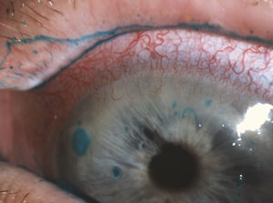
Corneal filaments stained with lissamine green are evident in this red, inflamed eye.
firmly adhere to compromised areas of the corneal epithelium, and a strand-like “tail” of varying length that extends across the ocular surface. Applying vital dyes, such as lissamine green, rose bengal and sodium fluorescein, can aid biomicroscopic filament visualization.3 A rapid tear break-up time and punctate epithelial keratopathy may also be present.
Not only does filamentary keratitis accompany dry eye, it also appears alongside a variety of other ocular surface disorders, including superior limbic keratoconjunctivitis, prolonged patching following ocular surgery, epitheliopathy due to aerosol or radiation keratitis, herpetic keratitis, recurrent corneal erosion, neurotrophic keratitis and bullous keratopathy.1-5
Mechanism Research suggests that individual filaments consist of desquamated corneal epithelial cells (at their cores), surrounded primarily by degenerating conjunctival epithelial cells and entwined in a thick layer of membrane-associated mucins.4,5 Patients with filamentary keratitis appear to suffer progressive dysfunction within the basal epithelial and Bowman’s layers of the cornea, leading to focal detachments at the level of the basement membrane. Under constant shear pressure from the eyelids, these corneal foci become inflamed, and sloughing of epithelial cells may ensue.6 At the same time, frictional stress from blinking and eye movement combined with diminished tear volume and ocular surface inflammation results in abnormal tear mucin production and degeneration of conjunctival epithelial cells.5  
These combined elements form filaments, which may appear clinically as long strands, large clumps or irregular dendriform deposits, depending upon whether they are stretched, twisted or tightly coiled.4,7 The filaments are motile in the tear film, but have an affinity for compromised areas of the corneal surface, where they form strong adhesions. Lid movement across these bound filaments induces vertical traction and further shearing of the corneal epithelium with each blink, resulting in microtrauma and stimulation of the pain-sensitive corneal nerves. Thus, a vicious cycle of epithelial damage, inflammation and filament formation ensues.
Management The management of filamentary keratitis is aimed at alleviating the stressors that cause ocular surface inflammation and epithelial degradation. Elimination of the filaments is the initial step, but identifying and treating the underlying pathology is vital to breaking the cycle of this disease. You can remove large filaments mechanically using fine-tipped forceps at the slit lamp under topical anesthesia. Recognize, however, that this process can further contribute to epithelial damage and should be undertaken only by skilled and experienced clinicians. Ocular lubricants are helpful in addressing discomfort and also stabilizing the tear film in mild to moderate cases. 
In recalcitrant cases, topical N-acetylcysteine can help to dissolve cornea-bound mucus plaques.1 This mucolytic agent is employed primarily as an oral inhalant for patients with bronchial disease (e.g. emphysema, cystic fibrosis), and hence it must be prepared by a compounding pharmacist for topical ophthalmic use. In those with filamentary keratitis secondary to chronic dry eye disease, we have seen excellent results with 10% acetylcysteine eye drops used four times daily for several weeks. Other treatments for refractory cases of filamentary keratitis may include the use of bandage soft contact lenses, amniotic membrane therapy or Botox (onabotulinumtoxinA, Allergan) injection to the pretarsal orbicularis muscle.2,8
Long-term Treatment Addressing the underlying ocular surface disease may ultimately prove more challenging than temporary elimination of corneal filaments. Because an inflammatory etiology is often assumed, the use of anti-inflammatory drugs such as corticosteroids and non-steroidal agents has been widely advocated, often with clinical success.9,10 In those cases where dry eye disease is identified as the primary etiology of filamentary keratitis, short-term use of corticosteroids such asLotemax (loteprednol etabonate 0.5%, Bausch + Lomb) QID combined with long-term use of Restasis (cyclosporine, Allergan) BID can help.11  
Severe cases may require treatment with autologous serum eye drops, which—as the name implies—are derived from the patient’s own blood serum.12,13 
Therapy for filamentary keratitis may take weeks or even months before adequate resolution is realized, depending on the etiology, severity of presentation and aggressiveness of care. Patients should understand that the underlying condition is often chronic and filaments may recur after therapy is discontinued. Proper long-term care includes ongoing treatment for ocular surface disease with close monitoring, i.e., three to four times annually. In addition, patients with chronic or severe dry eye disease may benefit from a rheumatologic investigation to determine the presence of Sjögren’s syndrome.14
Dr. Kabat is a consultant to Alcon Laboratories, Bio-Tissue and BlephEx. Neither he nor Dr. Sowka has any direct financial interest in the products mentioned in this article.

1. Albietz J, Sanfilippo P, Troutbeck R, Lenton LM. Management of filamentary keratitis associated with aqueous-deficient dry eye. Optom Vis Sci. 2003 Jun;80(6):420-30. 2. Gumus K, Lee S, Yen MT, Pflugfelder SC. Botulinum toxin injection for the management of refractory filamentary keratitis. Arch Ophthalmol. 2012 Apr;130(4):446-50.  3. Diller R, Sant S. A case report and review of filamentary keratitis. Optometry. 2005 Jan;76(1):30-6. 4. Tabery HM. Filamentary keratopathy: a non-contact photomicrographic in vivo study in the human cornea. Eur J Ophthalmol. 2003 Aug-Sep;13(7):599-605. 5. Tanioka H, Yokoi N, Komuro A, et al. Investigation of the corneal filament in filamentary keratitis. Invest Ophthalmol Vis Sci. 2009 Aug;50(8):3696-702. 6. Zaidman GW, Geeraets R, Paylor RR, Ferry AP. The histopathology of filamentary keratitis. Arch Ophthalmol. 1985 Aug;103(8):1178-81. 7. Pandit RT. Dendriform filamentary keratopathy. Cornea. 2009 Jan;28(1):123-5.  8. Suri K, Kosker M, Raber IM, et al. Sutureless amniotic membrane ProKera for ocular surface disorders: short-term results. Eye Contact Lens. 2013 Sep;39(5):341-7. 9. Perry HD, Doshi-Carnevale S, Donnenfeld ED, Kornstein HS. Topical cyclosporine A 0.5% as a possible new treatment for superior limbic keratoconjunctivitis. Ophthalmology. 2003 Aug;110(8):1578-81. 10. Terry G Coursey, Cintia S de Paiva. Managing Sjögren’s Syndrome and non-Sjögren Syndrome dry eye with anti-inflammatory therapy. Clin Ophthalmol. 2014; 8: 1447–1458. 11. Sheppard JD, Donnenfeld ED, Holland EJ, et al. Effect of loteprednol etabonate 0.5% on initiation of dry eye treatment with topical cyclosporine 0.05%. Eye Contact Lens. 2014 Sep;40(5):289-96. 12. Jirsova K, Brejchova K, Krabcova I, et al. The application of autologous serum eye drops in severe dry eye patients; subjective and objective parameters before and after treatment. Curr Eye Res. 2014 Jan;39(1):21-30.  13. Hussain M, Shtein RM, Sugar A, et al. Long-term use of autologous serum 50% eye drops for the treatment of dry eye disease. Cornea. 2014 Dec;33(12):1245-51.  14. Shen L, Kapsogeorgou EK, Yu M, et al. Evaluation of salivary gland protein 1 antibodies in patients with primary and secondary Sjogren’s syndrome. Clin Immunol. 2014 Nov;155(1):42-6.


Shopping Cart