There are many ways to diagnose papilledema but it can be tough to definitively confirm true papilledema versus pseudopapilledema without a lumbar puncture / spinal tap.
Here is the best information I could find.
1. papilledema is first diagnosed as suspicious based on the optic nerve exam under the slit lamp microscope in the majority of patients.
2. If it is not clear it is true papilledema, the following are recommended.
- Fluorescein angiography can detect disc leakage. Autofluorescence can demonstrate drusen and B scan ultraound may also be helpful to find drusen.
- Stereo disc images obtained to document and follow disc status.
- Perimetry commonly shows enlarged blind spots. In severe chronic papilledema peripheral field loss may be seen.
- CNS imaging study (CT or MRI with contrast) to identify a central nervous system mass lesion.
- MR venography should always be considered to look for venous sinus thrombosis.
- Lumbar puncture (after imaging demonstrates no risk for herniation) to document increased ICP and to look for neoplastic, infectious or inflammatory causes. At a minimum, opening pressure along with glucose, protein, cell count and differential, culture are obtained
Newer advanced MR imaging techniques such as fMRI and DTI may prove useful in the future to assess the potential effects of papilledema on retinal and visual pathway integrity.
Below is what the radiologist is looking for on the MRI.
MR Imaging Findings
A number of studies have used imaging techniques to investigate the anatomic changes of the ON in papilledema.5,6,8,9,12⇓⇓–15 Of the many imaging techniques, MR imaging has been of particular interest because of its ability to provide gross visualization of the optic globe, ON, orbits, and optic tract. Additionally, MR imaging provides higher soft-tissue contrast and free section orientation capabilities compared with CT and appears to be more accurate in assessing the ON than sonography.16
Despite these advantages, the ON has been technically difficult to image because of its small size: It is 0.4–0.6 cm in diameter within the orbit.8 T2-weighted FSE sequences with fat-suppression have been found to be optimal for visualizing the ONs and perioptic CSF.6,17⇓–19 Coronal image acquisition is optimal for visualizing the true dimensions of the ON and perioptic CSF relative to the surrounding sheath.12 The most commonly reported macroscopic findings in MR images of patients diagnosed with papilledema are the following: 1) enlargement of the ONS,5,6,8,9,20 2) flattening of the posterior sclera,13⇓–15 3) protrusion of the optic papilla into the globe,6,11 and 4) tortuosity of the ON.6
Enlargement of the ONS
The SAS around the ON in the orbit can be observed by using T2-weighted MR imaging with fat-saturation pulse sequences (Fig 1). The ONS diameter can be evaluated by measuring the outer diameter of the SAS.5
Fig 1.
ONS enlargement. A, Coronal T2-weighted FSE image demonstrates bilateral widening of the ONS in a 32-year-old woman who presented with acute vision loss and headaches and was found to have papilledema on fundoscopic examination. The ONSs are abnormally increased in size (TR, 6816.7 ms/TE, 84 ms; 3-mm thickness; FOV, 18 cm; matrix, 256 × 224; 4 excitations). B, Axial T2-weighted FSE image demonstrates the widening of the ONS bilaterally in a 31-year-old woman with known papilledema presenting with headaches (TR, 4400 ms/TE, 80 ms; 5-mm thickness; 6-mm spacing; FOV, 24 cm; 320 × 256 matrix).
The normal ONS diameters just behind and 4 mm posterior to the globe are 5.52 ± 1.11 and 5.2 ± 0.9 mm, respectively.5 The ONS is widest anteriorly behind the globe and narrowed toward the orbital apex8; these dimensions are consistent with the results of a histologic study of the ON.12 The sheath of the ON behind the globe is the most distensible part of the ONS, giving it a bulbous appearance.12
ONS enlargement appears as a widened ring of CSF around an ON and as a widened CSF signal intensity on either side of the ON on axial images.6 The finding of ONS enlargement have been reported previously in association with intracranial hypertension on sonography4 and CT.21 Seitz et al9 used MR imaging to examine patients with papilledema and found that the mean width of the ONS directly behind the globe was 7.54 mm (±1.05 mm) in pathologic conditions, compared with 5.52 mm (±1.11 mm) in healthy subjects. In addition, the CSF surrounding the ON from the optic globe toward the optic chiasm was visible over a longer distance (12.4 mm) in patients compared with healthy subjects (6.3 mm).9 Similar findings were noted in patients with IIH, with an enlarged and elongated SAS around the ON.8 The orbital portion of the ON was examined in patients at different stages of papilledema, and the diameter of the ONS was found to be increased compared with that in healthy subjects.20 With severe papilledema, the diameter of the ON just behind the globe was significantly less than that with moderate papilledema; this difference indicates that increased subarachnoid pressure may lead to gradual atrophy of the ON.20 Dilation of the SAS, compression of the ON, and widening of the ONS were also observed with optic disc pallor.12 Broadening of the ON directly behind the optic globe may also occur with papilledema.9
MR imaging has been used to evaluate the effectiveness of treating elevated ICP with ONS fenestration by assessing the resolution of the optic sheath enlargement.5,8 ICP and ONS diameter in patients with chronic subdural hematoma or hygroma who underwent burr-hole craniotomy and continuous drainage have shown significant correlation.5 ONS diameter before surgery (6.1 mm) was significantly reduced after surgery (4.8 mm).5 These findings suggest that the ONS diameter is a strong indicator of increased ICP.5,8
Flattening of the Posterior Sclera
Several studies have demonstrated posterior scleral flattening (Fig 2) in patients with elevated ICP.13⇓–15Gibby et al13 noted this finding on CT scans and considered it to be the mildest in the spectrum of changes leading to the protrusion of the ON head into the globe. Atta and Byrne14 noted flattening of the posterior sclera on sonography in some patients with choroidal folds and papilledema. Jacobson15 found bilateral flattening of the posterior sclera and distension of the perioptic SAS on MR imaging in a patient with elevated ICP and unilateral papilledema. These findings suggest that the combination of acquired hyperopia and choroidal folds may indicate pseudotumor cerebri in rare patients whose distal ONs are structurally resistant to developing papilledema.
Fig 2.
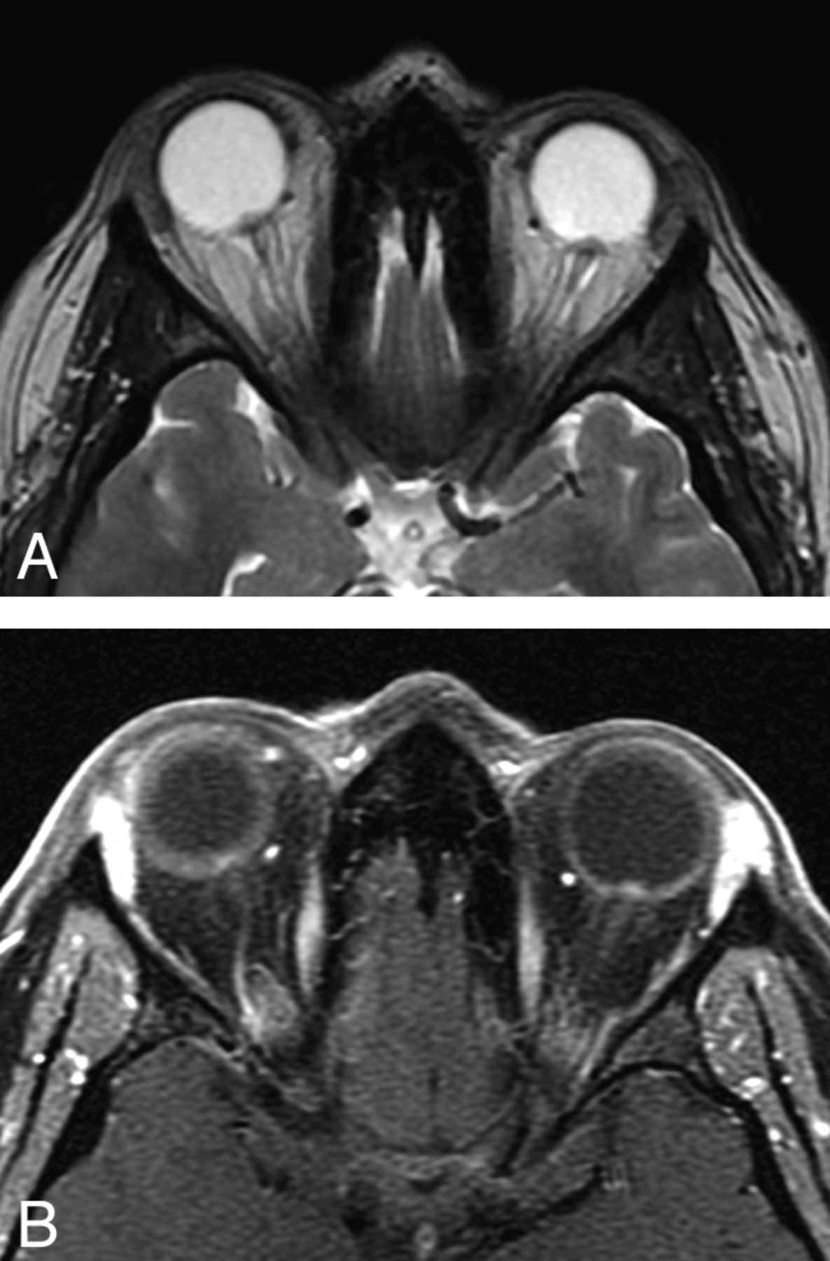
Optic papilla protrusion. In this T2-weighted axial image, both ON papillae protrude into the vitreous space of the globe. This 32-year-old woman had experienced headaches and vision loss on presentation; she was also found to have papilledema on fundoscopic examination (same patient as in Fig 1A). A, Axial T2-weighted image demonstrates bilateral ON head protrusion (TR, 3000 ms/TE, 84 ms; 5-mm thickness; FOV, 24 cm; matrix, 320 × 256; 1 excitation). B, Corresponding axial T1-weighted postcontrast image demonstrates enhancement of the ON heads (TR, 766.7 ms/TE, 9 ms; 3-mm thickness; FOV, 18 cm; matrix, 320 × 191; 1 excitation).
Protrusion of the Optic Papilla into the Globe
The optic papilla is considered by some to be the site most vulnerable to the effects of elevated CSF pressure in the nerve sheath.17 Normally, the optic papilla appears as a flat hyperintense (hypointense relative to the vitreous of the globe of the eye) region in the posterior sclera. In pseudotumor cerebri, intraocular protrusion of the optic papilla is occasionally observed on MR imaging. Brodsky and Vaphiades6 found prelaminar enhancement in 50% of MR images of patients with pseudotumor cerebri. However, they also found an absence of prelaminar enhancement in some patients with florid papilledema. Intraocular protrusion was also demonstrated by Jinkins et al11 in 10 (67%) of 15 patients with pseudotumor cerebri on MR imaging.
Intraocular protrusion of the optic disc (Fig 3) may not produce a sufficient signal-intensity differential with the vitreous of the globe on routine MR imaging. However, contrast-enhanced MR imaging may demonstrate a focal hyperintensity in the region of papilla protrusion because intraocular protrusion of the optic papilla disrupts blood flow in the optic prelaminar capillaries.6The optic papilla usually appears hypointense relative to the vitreous fluid of the globe on T2-weighted images because it is largely composed of myelinated axons. However, extracellular edema generally produces increased signal intensity on T2-weighted images, and this seems to contradict active capillary leakage. Histologic studies in patients with papilledema have demonstrated that despite capillary leakage, prelaminar axons appear distended but show little evidence of extracellular edema. This could explain the hypointensity of the optic papilla relative to the vitreous on T2-weighted images.11
Fig 3.
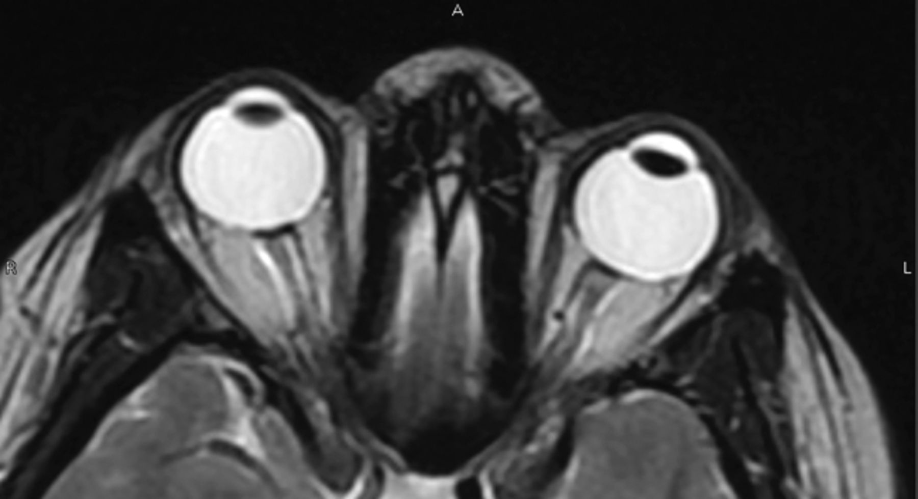
Posterior globe flattening. Flattening of the posterior globe is thought to occur in the same setting as papilledema with increased ICP. In this image, the site of the optic papilla is flattened and the normal globe contour is lost. This 37-year-old woman presented with headache and a sensation of head pressure. She was found to have papilledema and increased CSF opening pressure on lumbar puncture. (TR, 5650 ms/TE, 88 ms; 5-mm thickness; FOV, 24 cm; 320 × 256 matrix).
Tortuosity of the ON
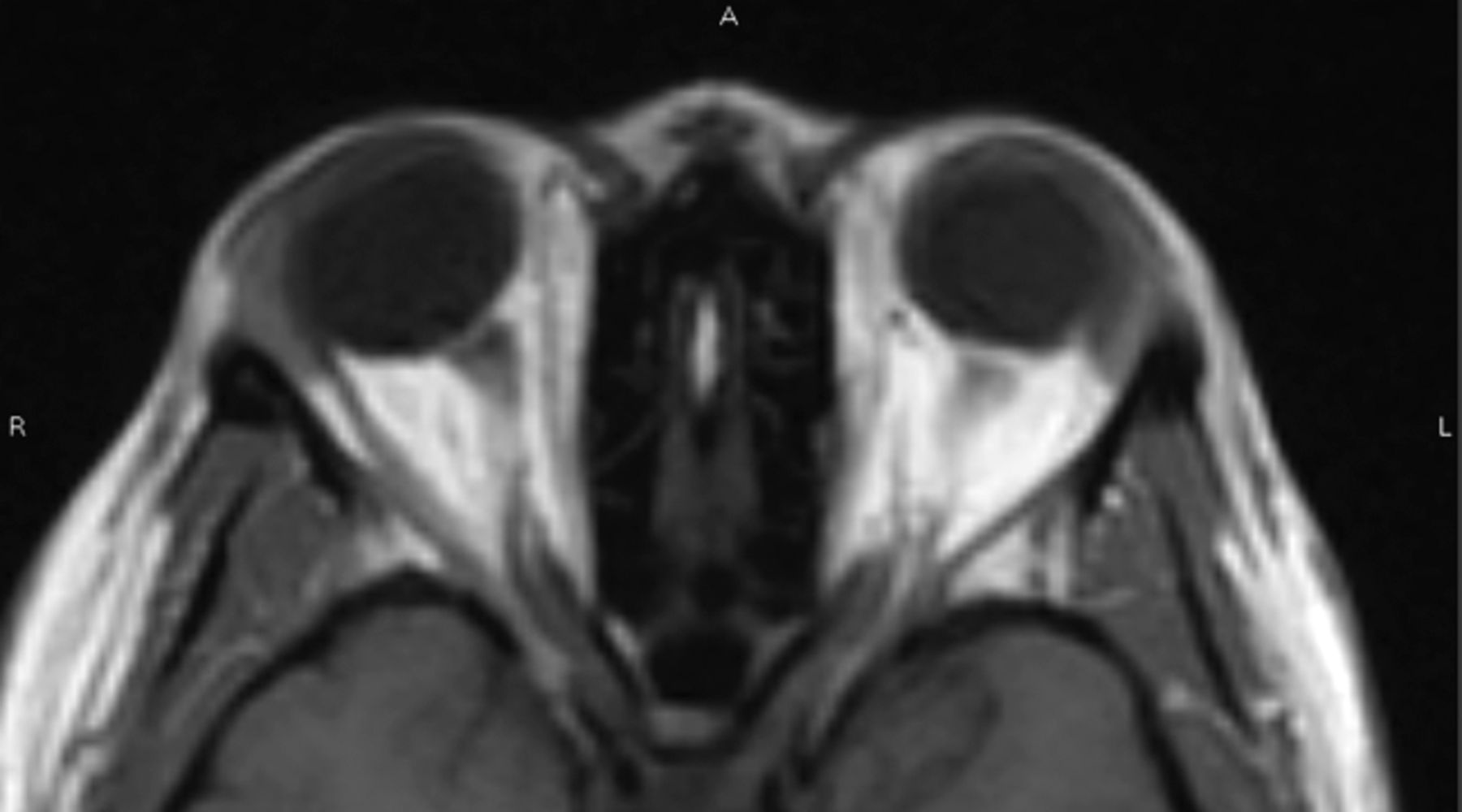
ON kinking or tortuosity (Fig 4) has also been associated with increased ICP. The ON tortuosity in patients with elevated ICP is attributable to the fixation of the distal and proximal points of the ON. The sensitivity of observing ON tortuosity in either the vertical or horizontal planes also depends on section thickness. The ability of axial MR imaging to display relatively minor degrees of horizontal tortuosity makes it a somewhat nonspecific finding. Tortuosity of the vertical component, which requires a greater deflection of the ON to be visible on axial scans, retains a higher specificity value. Vertical tortuosity of the ON is often accompanied by a “smear sign,” in which the midportion of the ON appears obscured by a “smear” of orbital fat on T1-weighted images (Fig 4).6
Fig 4.
ON tortuosity. Bending of the ON can be seen more prominently in the right ON in this 40-year-old woman with history of headache diagnosed with increased ICP. In this particular case, there is a smear sign, in which orbital fat obscures part of the tortuous ON (TR, 620 ms/TE, 9 ms/TI, 0 ms; 5-mm thickness; 6-mm spacing; FOV, 24 cm; 256 × 192 matrix).
Brodsky and Vaphiades6 theorized that perioptic distension would be expected to straighten the ONS if the globes were not tethered in the orbits by their rectus muscles and check ligaments. However, the absence of proptosis in patients with papilledema suggests that the focal pressure exerted by the bulbous portion of the distended perioptic nerve sheaths on the posterior sclera is insufficient to displace the globes anteriorly. Instead, the globe provides resistance to the distal bulbous portion of the ONS, which may kink the inflated ONS.6
MR Imaging Findings
A number of studies have used imaging techniques to investigate the anatomic changes of the ON in papilledema.5,6,8,9,12⇓⇓–15 Of the many imaging techniques, MR imaging has been of particular interest because of its ability to provide gross visualization of the optic globe, ON, orbits, and optic tract. Additionally, MR imaging provides higher soft-tissue contrast and free section orientation capabilities compared with CT and appears to be more accurate in assessing the ON than sonography.16
Despite these advantages, the ON has been technically difficult to image because of its small size: It is 0.4–0.6 cm in diameter within the orbit.8 T2-weighted FSE sequences with fat-suppression have been found to be optimal for visualizing the ONs and perioptic CSF.6,17⇓–19 Coronal image acquisition is optimal for visualizing the true dimensions of the ON and perioptic CSF relative to the surrounding sheath.12 The most commonly reported macroscopic findings in MR images of patients diagnosed with papilledema are the following: 1) enlargement of the ONS,5,6,8,9,20 2) flattening of the posterior sclera,13⇓–15 3) protrusion of the optic papilla into the globe,6,11 and 4) tortuosity of the ON.6
Enlargement of the ONS
The SAS around the ON in the orbit can be observed by using T2-weighted MR imaging with fat-saturation pulse sequences (Fig 1). The ONS diameter can be evaluated by measuring the outer diameter of the SAS.5
Fig 1.
ONS enlargement. A, Coronal T2-weighted FSE image demonstrates bilateral widening of the ONS in a 32-year-old woman who presented with acute vision loss and headaches and was found to have papilledema on fundoscopic examination. The ONSs are abnormally increased in size (TR, 6816.7 ms/TE, 84 ms; 3-mm thickness; FOV, 18 cm; matrix, 256 × 224; 4 excitations). B, Axial T2-weighted FSE image demonstrates the widening of the ONS bilaterally in a 31-year-old woman with known papilledema presenting with headaches (TR, 4400 ms/TE, 80 ms; 5-mm thickness; 6-mm spacing; FOV, 24 cm; 320 × 256 matrix).
The normal ONS diameters just behind and 4 mm posterior to the globe are 5.52 ± 1.11 and 5.2 ± 0.9 mm, respectively.5 The ONS is widest anteriorly behind the globe and narrowed toward the orbital apex8; these dimensions are consistent with the results of a histologic study of the ON.12 The sheath of the ON behind the globe is the most distensible part of the ONS, giving it a bulbous appearance.12
ONS enlargement appears as a widened ring of CSF around an ON and as a widened CSF signal intensity on either side of the ON on axial images.6 The finding of ONS enlargement have been reported previously in association with intracranial hypertension on sonography4 and CT.21 Seitz et al9 used MR imaging to examine patients with papilledema and found that the mean width of the ONS directly behind the globe was 7.54 mm (±1.05 mm) in pathologic conditions, compared with 5.52 mm (±1.11 mm) in healthy subjects. In addition, the CSF surrounding the ON from the optic globe toward the optic chiasm was visible over a longer distance (12.4 mm) in patients compared with healthy subjects (6.3 mm).9 Similar findings were noted in patients with IIH, with an enlarged and elongated SAS around the ON.8 The orbital portion of the ON was examined in patients at different stages of papilledema, and the diameter of the ONS was found to be increased compared with that in healthy subjects.20 With severe papilledema, the diameter of the ON just behind the globe was significantly less than that with moderate papilledema; this difference indicates that increased subarachnoid pressure may lead to gradual atrophy of the ON.20 Dilation of the SAS, compression of the ON, and widening of the ONS were also observed with optic disc pallor.12 Broadening of the ON directly behind the optic globe may also occur with papilledema.9
MR imaging has been used to evaluate the effectiveness of treating elevated ICP with ONS fenestration by assessing the resolution of the optic sheath enlargement.5,8 ICP and ONS diameter in patients with chronic subdural hematoma or hygroma who underwent burr-hole craniotomy and continuous drainage have shown significant correlation.5 ONS diameter before surgery (6.1 mm) was significantly reduced after surgery (4.8 mm).5 These findings suggest that the ONS diameter is a strong indicator of increased ICP.5,8
Flattening of the Posterior Sclera
Several studies have demonstrated posterior scleral flattening (Fig 2) in patients with elevated ICP.13⇓–15Gibby et al13 noted this finding on CT scans and considered it to be the mildest in the spectrum of changes leading to the protrusion of the ON head into the globe. Atta and Byrne14 noted flattening of the posterior sclera on sonography in some patients with choroidal folds and papilledema. Jacobson15 found bilateral flattening of the posterior sclera and distension of the perioptic SAS on MR imaging in a patient with elevated ICP and unilateral papilledema. These findings suggest that the combination of acquired hyperopia and choroidal folds may indicate pseudotumor cerebri in rare patients whose distal ONs are structurally resistant to developing papilledema.
Fig 2.
Optic papilla protrusion. In this T2-weighted axial image, both ON papillae protrude into the vitreous space of the globe. This 32-year-old woman had experienced headaches and vision loss on presentation; she was also found to have papilledema on fundoscopic examination (same patient as in Fig 1A). A, Axial T2-weighted image demonstrates bilateral ON head protrusion (TR, 3000 ms/TE, 84 ms; 5-mm thickness; FOV, 24 cm; matrix, 320 × 256; 1 excitation). B, Corresponding axial T1-weighted postcontrast image demonstrates enhancement of the ON heads (TR, 766.7 ms/TE, 9 ms; 3-mm thickness; FOV, 18 cm; matrix, 320 × 191; 1 excitation).
Protrusion of the Optic Papilla into the Globe
The optic papilla is considered by some to be the site most vulnerable to the effects of elevated CSF pressure in the nerve sheath.17 Normally, the optic papilla appears as a flat hyperintense (hypointense relative to the vitreous of the globe of the eye) region in the posterior sclera. In pseudotumor cerebri, intraocular protrusion of the optic papilla is occasionally observed on MR imaging. Brodsky and Vaphiades6 found prelaminar enhancement in 50% of MR images of patients with pseudotumor cerebri. However, they also found an absence of prelaminar enhancement in some patients with florid papilledema. Intraocular protrusion was also demonstrated by Jinkins et al11 in 10 (67%) of 15 patients with pseudotumor cerebri on MR imaging.
Intraocular protrusion of the optic disc (Fig 3) may not produce a sufficient signal-intensity differential with the vitreous of the globe on routine MR imaging. However, contrast-enhanced MR imaging may demonstrate a focal hyperintensity in the region of papilla protrusion because intraocular protrusion of the optic papilla disrupts blood flow in the optic prelaminar capillaries.6The optic papilla usually appears hypointense relative to the vitreous fluid of the globe on T2-weighted images because it is largely composed of myelinated axons. However, extracellular edema generally produces increased signal intensity on T2-weighted images, and this seems to contradict active capillary leakage. Histologic studies in patients with papilledema have demonstrated that despite capillary leakage, prelaminar axons appear distended but show little evidence of extracellular edema. This could explain the hypointensity of the optic papilla relative to the vitreous on T2-weighted images.11
Fig 3.
Posterior globe flattening. Flattening of the posterior globe is thought to occur in the same setting as papilledema with increased ICP. In this image, the site of the optic papilla is flattened and the normal globe contour is lost. This 37-year-old woman presented with headache and a sensation of head pressure. She was found to have papilledema and increased CSF opening pressure on lumbar puncture. (TR, 5650 ms/TE, 88 ms; 5-mm thickness; FOV, 24 cm; 320 × 256 matrix).


