Cold Eeze and Alzheimer’s Disease

There are no studies to date linking Cold Eeze and Alzheimer’s Disease. Still I am concerned about the long term consequences of using Zinc lozenges to fight a cold. I love Cold Eeze and I use it many times a year. I know it works for me to prevent colds.

Yet, there are some studies linking Zinc oxide to Alzheimers and I am not sure how much Zinc Gluconium (the key ingredient in Cold Eeze) translates into Zinc oxide flowing through my brain. Am I putting myself at risk  by taking excessive amounts of zince 4-5 times per year?

Maybe.

Each Cold Eeze has 13.3mg of Zinc Gluconium. The upper limit recommended of Zinc is 40mg. The Cold Eeze box says use every 2-4 hrs until symptoms subside. If we assume I take it every 2 hrs, which I have done in the past, even sleeping with one in my mouth, which I definitely do NOT recommend (but I was desperate to not get sick), then my total for a 24hr period is 159.6mg. This is excessive.

Here are some recent studies looking into the role of Zinc in Alzheimers.

For now the key is to avoid going over 40mg per day. But will Cold Eeze truly work for me in this way? That is only 3 Cold Eeze a day. I will find out, but I am suspicious I need more Zinc lozenges to fight a cold. I still follow the bone broth/chicken soup intake I recommended in a previous post.

https://drcremers.com/2017/05/best-ways-to-fight-cold-and-from.html
And hot showers, massaging, etc. But will that be enough to fight a cold without excessive Cold Eeze?

To be continued.
SLC

. 2018; 8: 4772.
Published online 2018 Mar 19. doi:  10.1038/s41598-018-23122-x
PMCID: PMC5859292
PMID: 29555950

Zinc ion rapidly induces toxic, off-pathway amyloid-β oligomers distinct from amyloid-β derived diffusible ligands in Alzheimer’s disease

Open Access This article is licensed under a Creative Commons Attribution 4.0 International License, which permits use, sharing, adaptation, distribution and reproduction in any medium or format, as long as you give appropriate credit to the original author(s) and the source, provide a link to the Creative Commons license, and indicate if changes were made. The images or other third party material in this article are included in the article’s Creative Commons license, unless indicated otherwise in a credit line to the material. If material is not included in the article’s Creative Commons license and your intended use is not permitted by statutory regulation or exceeds the permitted use, you will need to obtain permission directly from the copyright holder. To view a copy of this license, visit http://creativecommons.org/licenses/by/4.0/.

Introduction

AD is the most common cause of dementia in the elder population after age of 65. All current AD clinical trials have failed due to insignificant beneficial effects or severe adverse effects,. The failure of clinical trials suggests that the fundamental molecular mechanism of AD pathogenesis is still not fully understood. Aβ, a pathogenic hallmark in AD, is cleaved from amyloid precursor protein by β- and γ-secretases,. Aβ40 and Aβ42 are the two major isoforms that differ in two additional amino acids in the C- terminus of Aβ,. Aβ is an intrinsically disordered protein that is prone to aggregate into cross-β-rich fibrils via a nucleation-dependent manner. The classic amyloid fibrillization consists of a nucleation state followed by fibril elongation and a plateau for mature fibril formation. The major cause of AD is considered to associate with assemble of Aβ into oligomers, which impair synaptic function and lead to activation of a cascade of subsequent detrimental events,. Aβ oligomers are previously referred to heterogeneous intermediates in the aggregation, including various types of species, e.g. prefibrillar oligomer, protofibrils, annular protofibrils, paranuclei, globulomers, amylospheroids, ADDLs, and Aβ56*. Despite the intrinsic structural heterogeneity of Aβ oligomeric aggregates, many of their structural features have been unraveled by solid-state NMR. Aβ fibrillization can be monitored by thioflavin T (ThT) that emits fluorescence upon chelating to cross-β-stands in amyloid fibrils, however, the oligomer intermediates showed no or low binding to ThT.
Although Aβ40 and Aβ42 are the two major Aβ isoforms, they have distinct properties in structure, aggregation, and toxicity. Freshly prepared Aβ40 was reported to be monomer and Aβ42 adopts rapid equilibrium of monomer and trimer/tetramer. Aβ40 and Aβ42 adopt distinct fibrillization pathways,. During the aggregation, Aβ42 forms a pentameric/hexameric paranuclei, whereas, Aβ40 undergoes monomer addition,. Ion mobility mass spectrometry showed Aβ40 assembles through tetramer, whereas, Aβ42 forms tetramer and further forms hexamer that stacks into dodecamer prior to protofibril/fibril formation. The fibril structure of Aβ40 contains two β-strands formed by amino acids 10 to 22 and 30 to 40 and Aβ42 fibrils contain multiple β-sheets adopting the so-called LS-shaped structure. Aβ42 is demonstrated more detrimental than Aβ40 in vitro and in vivo, despite the amount of Aβ42 is 10 times less than Aβ40 in cerebrospinal fluids.
Metal ion dyshomeostasis is an existing concept in the AD research. Abnormally high content of metal ions including Zn2+, Cu2+, Fe3+ have been found in senile plaques of postmortem AD brains,. Many evidences further suggested imbalance of Zn2+ or Cu2+ homeostasis was linked with AD pathology,and association of metal ions with Aβ, especially Cu2+ or Zn2+ were reported in a series of studies. Aβ is known to bind with the metal ions,. The metal ions, including Zn2+ and Cu2+, were identified to interact with histidine residues at N-terminal of Aβ,. The mechanism of interaction of Zn2+ with Aβ was reported to share similar coordination with Cu2+ by three histidines at residue 6, 13, and 14 of Aβ,. However, how Zn2+ modulates Aβ conformation is still elusive, that may be due to assemblies of ZnAβ were variable and sensitive due to changes of pH, temperature, concentration, and buffer environment. The morphology of ZnAβ was previously shown as fibrils at 37 °C, whereas others discovered that ZnAβ formed amorphous aggregates or oligomers at room temperature,. Postmortem brain analysis and in vivo model of AD both provided evidences to support that Zn2+ might play a role in AD pathologensis,. Bush et al. further developed a Cu/Zn ion chelator, PBT2, to provide beneficial effect for AD,.
To understand the effect of Zn2+ to Aβ40 and Aβ42, here we systematically characterized the oligomer formed by ZnAβ complex. We found Zn2+ rapidly induced secondary structure changes and retained Aβ in an oligomer state with higher hydrophobic surface. ZnAβ oligomers have distinct immunoreactivity against several anti-Aβ antibodies and possess higher toxicity than ADDLs. Removal of Zn2+ leads to restoration of Aβ fibrillization with a faster kinetics than Aβ alone. In addition, solid-state NMR data suggested that ZnAβ40 oligomers possess β-sheet structure. The electrophysiology result showed that ZnAβ oligomer inhibited hippocampal LTP in the wild-type mice hippocampus slice. In the acute Aβ-injected mice model, increasing of hippocampal microglial activation was found followed by ZnAβ injection.

Results

Zn2+ promotes Aβ40 and Aβ42 oligomer formation

To systematically examine the effect of Zn2+ on Aβ aggregation, we incubated equal molar ratio of Zn2+ions with Aβ40 and Aβ42 and examined their fibrillization by ThT assay, the secondary structural changes by far-UV CD, and the epitope changes of the assembly by dot blotting. Aβ40 and Aβ42 were prepared in ammonium hydroxide, lyophilized, and then dissolved in 10 mM Tris-HCl buffer, pH 7.4. Fifty µM Aβ and Zn2+ were used in the experiments. In ThT assay, we found that in the absence of Zn2+, Aβ40 fibrillization adapted a classic nucleation-dependent response. It showed a lag phase of ~64 hr, and reached maximal ThT fluorescence at ~94 hr (Fig. 1A). Upon addition of equal molar concentration of Zn2+ during Aβ40 aggregation, we found the ThT signal was retained without much increase. The result showed that there is lower incidence, if any, of amyloid fibril formation in the presence of Zn2+ ions. In the case of Aβ42, Aβ42 alone showed a slow growing phase of ~20 hr, then increased to maximal ThT fluorescence intensity at ~33 hr (Fig. 1F). Whereas, in the presence of Zn2+, ThT fluorescence was significantly inhibited similar to the result obtained from Aβ40. The result demonstrated that Zn2+ suppresses both Aβ40 and Aβ42 fibrillization. We also examined Aβ40 and Aβ42 fibrillization in various concentrations of Zn2+ in ThT assay. Five different concentrations of Zn2+ ranging from 10 to 200 µM were added into the 50 µM Aβ solution. The result showed Zn2+ suppressed Aβ40 and Aβ42 fibrillization in a dose-dependent manner (Fig. S1). To examine the possible precipitation, we examined the turbidity of the samples for 7 days of incubation by absorbance at 340 nm and also quantified the protein concentration at 280 nm. The samples were turbid especially for the ZnAβ samples indicating large soluble aggregate formation, but no precipitation was observed during the experimental time (Fig. S2). To see whether there is any Aβ aggregation in the presence of Zn2+, we imaged the ZnAβ40 and ZnAβ42 aggregates by TEM (Fig. 1B,G, respectively). TEM images showed that in the absence of Zn2+ the end-point products of Aβ40 and Aβ42 both formed straight or curvy amyloid fibrils. In contrast, equal molar ratio of Zn2+ promoted formation of spherical Aβ species, whose diameters in average were ~14 and ~12 nm for ZnAβ40 and ZnAβ42, respectively, as measured from the TEM images (Fig. S3). Furthermore, we added equal amount of Zn2+into preformed Aβ40 fibrils and studied whether Zn2+ is capable to destabilize preformed fibrils by ThT assay and Fourier transform infrared spectra (FTIR). The results showed that the addition of Zn2+ ions cannot decrease ThT intensity of Aβ fibrils (Fig. S4A) and does not alter β-sheet structure signals at 1,630 cm−1 in FTIR analysis (Fig. S4B). This result suggested that Zn2+ only affects Aβ at the early stage.

Zn2+ promotes Aβ oligomerization. (A,F) ThT assay of Aβ in the absence or presence of Zn2+. Aβ or Aβ42 (F) at 50 µM was incubated with and without equal molar concentration of Zn2+. The final ThT concentration in the working solution is 5 µM. ThT fluorescence was monitored at 25 °C. (B,G) TEM images of the end-point products of Aβ alone or ZnAβ. The scale bars are 100 nm. (CE) and (HJ) Far-UV CD spectra of Aβ40 and Aβ42. The spectra were scanned from 250 to 190 nm at the indicated time. Aβ40 in the absence (C) and presence of Zn2+ (D) after different incubation time are shown. The single wavelength changes of Aβ40 at 200 and 220 nm after addition of Zn2+ are shown in panel (E). Aβ42 in the absence (H) and presence of Zn2+ (I) were also monitored. The single wavelength changes of Aβ42 at 200 and 220 nm after addition of Zn2+ were shown in panel (J).

Zn2+ rapidly changes secondary structures of Aβ40 and Aβ42 and retains Aβ in a less β-conformation

To understand how Zn2+ affects the conformation of Aβ, the secondary structure of Aβ40 and Aβ42 with and without Zn2+ was examined by far-UV CD spectroscopy for several days (Fig. 1C–E, H–J). On day 0, freshly-prepared A40 showed a random-coil-like spectrum and the conformation gradually transformed to α-helix-like spectra on day 4 and 5, then finally formed β-sheet-rich structures which minimum wavelength at 217 nm on day 6 (Fig. 1C). For the spectra of Aβ42, the random-coil-like conformation on day 0 quickly moved to β-sheet-rich structure after day 1 (Fig. 1H), this result is consistent with the result of ThT assay showing Aβ42 forms fibrils much faster than Aβ40. When Zn2+ was mixed with Aβ40 and Aβ42, the ellipticity was significantly reduced on and after day 0 (Fig. 1D,I). The result showed that Zn2+ affects the secondary structure of Aβ rapidly and the aggregates contain different secondary structures than Aβ alone. Previously, this reduction was also found in the presence of Al3+ and other metal ions in our previous findings with a different Aβ preparation method. Interestingly, we found Zn2+ affects Aβ40 much more than Aβ42. The CD spectra of Aβ40 with Zn2+ did not show much secondary structure from day 1 to even after 6 days of incubation, whereas Aβ40 without Zn2+ formed β-sheet fibrils readily. In the presence of Zn2+ with Aβ42, the random-coil-like structure was also reduced at day 0 and transformed to mixed spectra after 1 day (Fig. 1I). On day 2, there is no obvious β-stand formation unlike the Aβ42 alone (Fig. 1H). The single wavelength changes of Aβ40 (Fig. 1E) and Aβ42 (Fig. 1J) at 200 and 220 nm are plotted against time after the addition of Zn2+. The results showed that Zn2+-induced Aβ changes were rapid and complete in minutes.

ZnAβ40 and ZnAβ42 form oligomers that are less heterogeneous and possess higher hydrophobic-exposed surfaces than ADDLs

To estimate the size of ZnAβ oligomers, we analyzed ZnAβ40 and ZnAβ42 oligomers and ADDLs composed of Aβ40 or Aβ42 by sedimentation velocity (SV) in analytical ultracentrifugation (AUC). ADDL is a type of Aβ oligomers commonly reported in the literature that are cytotoxic,,. ADDL was prepared as described in method according to previous literatures,,. The SV result showed that the predominant sedimentation coefficient distribution of ZnAβ40 or ZnAβ42 were similar. They ranged from 10 to 17.5 S corresponding to molecular weight ranging from 152 to 214 kDa (~33 to 49 mer). The peak maxima were situated at 11.3 and 11.9 S, respectively, where the peaks contained ~63% and ~46.2% of total Aβ (Fig. 2A,B). In the condition to form ADDLs by Aβ40, the major distribution were broadly distributed which indicates no distinct oligomer formed in this condition. This result is consistent with the previously report that no Aβ40 oligomer was observed under AFM in ADDL preparation. In ADDL made by Aβ42, there is a major peak situated at ~10.27 S which corresponds to molecular weight of 159 kDa (~35 mer), containing ~26.09% of total peptide. There are some other assemblies presented under 5 S which corresponds to molecular weight less than ~95 kDa (~21 mer). The friction ratio of ZnAβ40 and ZnAβ42 are 1.4 and 1.2, whereas, ADDL40 and ADDL42 are 1.57 and 1.48, respectively. Since the perfect sphere has a fraction ratio of 1.2, the result indicates that ZnAβ complexes are more spherical. Our AUC data demonstrated that ZnAβ40 and ZnAβ42 form oligomers that are less heterogeneous and more spherical than ADDLs.

ZnAβ40 or ZnAβ42 assemble into oligomers and contain more exposed hydrophobic surfaces than ADDLs. (A,B) SV in AUC of ZnAβ or ADDLs consist of Aβ40 or Aβ42, respectively. The data were analyzed by a continuous c(s) distribution model and the sedimentation coefficients were calculated and shown. (C,D) Bis-ANS spectra of ZnAβ and ADDL. ZnAβ40, ZnAβ42, ADDL-Aβ40, and ADDL-Aβ42 at 100 µM were prepared. Before the measurement, all Aβ solution were diluted to 50 µM in 10 mM Tris-HCl, pH 7.4, then, mixed with 0.5 µM Bis-ANS dye. Bis-ANS fluorescence spectra were collected. The spectra were subtracted from the respective buffer control.
Besides AUC analysis, we employed Bis-ANS fluorescence to monitor the hydrophobic clusters exposed on Aβ surface,,. The experiments were performed with 50 µM ZnAβ oligomers or ADDLs. The samples were mixed with Bis-ANS readily within 1 min and subjected to fluorescence measurement. The fluorescence spectra of Aβ in the presence of Zn2+ ion was shown to be ~2.8 and ~1.6 fold higher than that of ADDLs Aβ40 or Aβ42, respectively (Fig. 2C,D). This enhancement indicated higher level of hydrophobic surface exposed on the protein surface of ZnAβ oligomers. Besides increasing of fluorescence intensity, the spectra of ZnAβ40 oligomers also showed blue-shift of maximal wavelength from 499 nm to 485 nm, but the shift in the ZnAβ42 is not that obvious, indicating that the presence of Zn2+ altered the Bis-ANS binding environment.

Low immunoreactivity of ZnAβ oligomers reveal distinct oligomeric structure

To further elucidate the effect of Zn2+ on conformation changes of Aβ, we collected time-course Aβ aggregates for dot blotting analysis to monitor epitope changes (Fig. S5). Aβ40 and Aβ42 samples at different time points of incubation were dotted on the nitrocellulose membrane with several replicates and probed by several Aβ antibodies including conformational specific antibodies A11 and OC and sequence specific antibodies like 6E10, 4G8, A3356, and C-terminal antibody. A11 antibody was used to detect common prefibrillar oligomers, whereas OC antibody recognizes fibrils and fibrillar oligomers, which are structurally and immunologically related to fibrils,. The sequence specific antibodies used were 6E10 antibody recognizing N-terminal Aβ residues 1–16, 4G8 recognizing Aβ residues 17–24, A3356 recognizing the middle terminus of Aβ residues 22–35, anti-C-terminal of 42 recognizing the last 6 residues of Aβ42. The peptide loading control is detected by Direct Blue staining. From the different time points collected during Aβ40 and Aβ42 aggregation incubated with or without Zn2+ (Fig. S3A and S3B), we found A11 signals of Aβ40 increased after day 1, and that of ZnAβ40 increased after day 5. Both of the signal saturated after 6 days. In the OC blot, we found the signals of Aβ40 potently increased after day 1, but ZnAβ40 showed almost no OC signal in comparison to Aβ40 alone. Similarly, A11 and OC signals were both much weaker in the ZnAβ42 blot compared with Aβ42 alone. These evidences suggest that ZnAβ oligomers revealed distinct immunological status compared with Aβ aggregates in the absence of Zn2+. The data are consistent with the results from ThT assay and TEM imaging demonstrating no or little fibrillary structure is formed in ZnAβ. Surprisingly, we found that even sequence-dependent antibodies such as 6E10 or 4G8 showed weaker reactivity to recognize ZnAβ40 and ZnAβ42 while we have equal Aβ loading on the blot as shown by Direct Blue staining. Furthermore, we compared the epitopes in ZnAβ oligomers and ADDLs by the different antibodies. We dotted ZnAβ incubated for 1 and 7 days with ADDLs that were prepared in media for 1 day as described in the method. We found that both ZnAβ40 and ZnAβ42 incubated for 1 or 7 days showed weaker immunoreactivity compared with the corresponding ADDLs (Fig. S5C and S5D). This result further demonstrated that ZnAβ oligomers expose distinct epitopes and maintain a quite different conformation than ADDLs and the intermediates in Aβ aggregation without Zn2+.

ZnAβ40 oligomers are structurally more disordered than Aβ40 fibrils

It is well known that the chemical shifts of CO, Cα, and Cβ are sensitive to the backbone conformations of polypeptides. To understand the molecular structure of ZnAβ, we first examined the 13C uniformly labeled ZnAβ40 by solid-state homonuclear 13C correlation spectrum. In contrast to the highly resolved NMR spectra of Aβ40 fibrils, the resolution of the spectrum acquired for our ZnAβ40 sample was very poor (Fig. S6). It implied that ZnAβ40 still exhibited a significant structural heterogeneity at the molecular level. This finding suggested that the formation of ZnAβ40 aggregates is a kinetically controlled event. To tackle the resolution problems, we prepared a selectively labeled ZnAβ40 sample in which only the residues of V24, A30, I31, G33, and L34 were 13C labeled. The corresponding NMR spectrum is shown in Fig. 3. The assignments of the ZnAβ40 spectrum are given in the Supporting Information (Fig. S7). Accordingly, most of the cross peaks of V24 were significantly attenuated and only the Cβ/Cγ cross peak was barely observed. The Cα/Cβ cross peak of A30 was discernible but that of I31 was not observed. Nonetheless, the cross peaks of the side chain carbons of I31 remained observable. The CO/Cα cross peak of G33 were observed but its line width was relatively large, rendering an accurate determination of its chemical shifts difficult. The cross peaks of L34 were the most intense in the ZnAβ40 spectrum and there were at least two sets of resonances. For comparison, the labeled peptides were also incubated under quiescence conditions to form Aβ40 fibrils. Figure 3 showed the overlay 13C NMR spectrum of ZnAβ40 and Aβ40 fibrils. The chemical shifts and line widths of the backbone carbons of L34 were rather similar between ZnAβ40 and Aβ40 fibrils. On the other hand, the chemical shifts of ZnAβ40 revealed that the residue A30 is largely in the β-sheet conformation, but to a lower extent than Aβ40 fibrils. In addition, the line widths of the A30 signals of ZnAβ40 were larger than those of Aβ40.

Overlaid 13C homonuclear correlation spectra of ZnAβ40 (green) and Aβ40 fibrils (red). The Aβ40 peptides were 13C enriched at V24, A30, I31, G33, and L34. Spectral assignments were given for the spectrum of the Aβ40 fibrils. The dashed elliptical circle highlighted the region of the Cα/Cβ cross peaks of V24 and I31 expected for ZnAβ40. The peaks denoted by asterisks were spinning sidebands.

Removal of Zn2+ restores amyloid fibril formation with a faster kinetics and ZnAβ is unable to serve as fibrillization seeds

Since the presence of Zn2+ promoted Aβ40 or Aβ42 forming stable oligomers, we wonder whether metal ion chelator, such as EDTA, can disrupt the interaction of Zn2+ ions and Aβ. Here, we added three-fold molar concentration of EDTA into the ZnAβ or Aβ solution after plateau phase of ThT intensity at the time point indicated by arrows and monitored the conformation and morphology by far-UV CD and TEM image (Fig. 4). Aβ40 or Aβ42 in the absence or presence of Zn2+ were incubated and monitored by ThT assay (Fig. 4A,E). When the ThT intensity of Aβ alone reached a plateau phase, EDTA were added to both solution. We found that upon adding EDTA to trap Zn2+ ions, ZnAβ40 showed a rapid restoration to form amyloid fibrils with a short lag phase (<~40 hr), whereas ThT intensity of Aβ40 alone was not changed after EDTA addition (Fig. 4A). We also monitored the secondary structural changes by time-course far-UV CD spectra (Fig. 4B). Interestingly, the result showed that removal of Zn2+ ions immediately promoted ZnAβ40 to again show random-coil-like structure (labeled as day 0) similar to the Aβ40 monomer solution without Zn2+. The initial spectra of ZnAβ before addition of EDTA was flat (labeled as dash line). After EDTA treatment, Aβ gradually transformed to β-sheet-rich structure after 4 days of incubation. This transition is similar to conformational transition (6 days) in Aβ alone but faster. We monitored the morphology of the end-point products by TEM (Fig. 4C) and found ZnAβ40 after EDTA treatment has formed mature fibrils similar to the control Aβ without Zn2+. Dot blotting by A11 and OC antibody of the time course ZnAβ40 samples after EDTA treatment also demonstrated that trapping of Zn2+ by EDTA results in restoration of Aβ40 to fibrillization pathway (Fig. 4D). In the case of Aβ42, ThT assay showed that EDTA treatment also promoted ZnAβ42 to immediate formation of amyloid fibrils without a lag phase (Fig. 4E). However, in far-UV CD analysis, we found the spectra removal of Zn2+ ions by EDTA still showed the β-strand content which may be due to unable to monitor the rapid aggregation of Aβ42. Dot blotting and TEM further confirmed the restoration of ZnAβ42 into amyloid fibrils and restoration of the immunoreactivity (Fig. 4G,H).

ZnAβ40 oligomer can be restored rapidly back to fibrillization pathway by EDTA. (A,E) Aβ and ZnAβ40 (A) or ZnAβ42 (E) aggregation monitored by ThT assay. Three folds of EDTA was added when Aβ fibrillization reached a plateau. The time of addition is indicated by arrows. (B,F) Time-course conformation changes of ZnAβ40 or ZnAβ42 after EDTA treatment measured by far-UV CD. (C,D and G,H) TEM images (C and G) and dot blotting (D,H) of the time course samples in the aggregation. Scale bars in TEM images are 100 nm. ZnAβ was first prepared and incubated for 1 day for ZnAβ oligomer formation prior to EDTA treatment in the CD and dot blotting experiments. Dot blotted membrane was probed with A11 and OC antibody and stained by Direct blue as a dotting control.
Next, we would like to examine the property of Aβ-EDTA and ZnAβ-EDTA being seeded. We performed seeding experiment by addition of fibril seeds generated from preformed Aβ fibrils to soluble Aβ-EDTA and ZnAβ-EDTA solution and monitored the fibrillization kinetics by ThT fluorescence (Fig. 5A,B). The preformed Aβ40 or Aβ42 fibril seeds were sonicated and added in 5% to respective soluble Aβ solution. The result showed that all Aβ-EDTA and ZnAβ-EDTA can be seeded by fibril seeds showing accelerated fibrillization. There is no difference in initial kinetic rate of fibrillization, but the plateau phase of ThT intensity of ZnAβ-EDTA was markedly lower than Aβ-EDTA. This may indicate a distinct ThT binding environment or fibril structure formed from ZnAβ-EDTA fibrillization. Furthermore, we directly compared the fibrillization kinetics of Aβ and Zn-Aβ both treated with EDTA (Aβ-EDTA and ZnAβ-EDTA) by ThT assay (Fig. 5C,D). The result showed that the aggregation kinetics of ZnAβ40-EDTA was notably faster than Aβ40-EDTA with a shorter lag phase (ZnAβ40-EDTA, lag time = 33 hr; Aβ40-EDTA, lag time = 42 hr) (Fig. 5C), whereas, ZnAβ42-EDTA still showed similar kinetic profile as Aβ42-EDTA alone (Fig. 5D). The plateau phase of ThT intensity of both ZnAβ40-EDTA and ZnAβ42-EDTA were markedly lower than the respective Aβ-EDTA. The result is consistent with observing faster aggregation after Zn removal in aforementioned experiments (Fig. 4A,B). It suggests ZnAβ oligomer may facilitate formation of an Aβ conformation that is less heterogeneous and can facilitate the Aβ fibrillization after removal of Zn2+. However, the effect is not easily seen in Aβ42 that may be due to fast aggregation of Aβ42 hindering the observation of the difference.

EDTA-treated ZnAβ40 showed faster fibrillization rate and ZnAβ oligomers are unable to seed Aβ. (A,B) Fibrillization kinetics of (A) EDTA-treated ZnAβ40 or Aβ40 and (B) EDTA-treated ZnAβ42 or Aβ42 after seeding. Five % sonicated preformed fibril seeds were added into EDTA-treated ZnAβ and Aβ solution. Fibrillization kinetic was measured by ThT fluorescence. ZnAβ alone and buffer containing 5% fibril seeds were also monitored. (C,D) Fibrillization kinetics of Aβ40 (C) and Aβ42 (D) with or without Zn2+ after EDTA treatment. ZnAβ was first prepared and incubate for 1 day for oligomerization, then added EDTA to remove Zn2+ ion. Aβ alone solution was also treated with EDTA for comparison. Fibrillization kinetics was measured by ThT fluorescence. (E,F) Seeding experiment were performed by adding Aβ fibrils or ZnAβ40 (E) or ZnAβ42 (F) as seeds. The respective buffer were added to Aβ solution as negative control.
We further seeded preformed Aβ fibrils and ZnAβ oligomers to Aβ solution to examine the seeding property of ZnAβ in comparison to Aβ fibrils (Fig. 5E,F). Aβ40 and Aβ42 fibril seeds or ZnAβ40 and ZnAβ42 seeds were add 5% to the Aβ solution. The corresponding buffers were also added separately as negative controls. We found that Aβ fibril seeds can greatly accelerate fibrillization as expected. In Aβ40, the ThT signal of Aβ with fibril seeds elongated immediately without a lag phase compared to Aβ without fibril seeds. Whereas, Aβ with ZnAβ40 seeds did not show acceleration but adopted a similar aggregation characteristic of Aβ without seeds (buffer added). Similarly, in Aβ42, Aβ42 with Aβ42 fibril seeds adopted a faster elongation rate in comparison to the Aβ42 without seeds. ZnAβ42 oligomer did not seed Aβ42 solution. The results demonstrate ZnAβ cannot serve as seeds to accelerate fibrillization. Overall, the seeding experiments showed consistent results to indicate off-pathway property of ZnAβ.

ZnAβ oligomers possess higher cytotoxicity than ADDLs

Aβ oligomers have been proposed to be the major cause contributed to neurotoxicity in the Aβ assembly. Here, to understand whether ZnAβ oligomers show difference in the biological effects compared with ADDLs, we examined the cell viability and cytotoxicity contributed from ZnAβ oligomers and ADDLs. Since high Zn2+ ion concentration induces cell death,, we exchanged excess Zn2+ in the preparation with 10 mM Tris-HCl buffer, pH 7.4 by MWCO 3 KDa centrifugal filter and re-quantify concentration prior to further test. The remaining Zn concentration after exchanged was 2 µM (data not shown). The cell viability and cytotoxicity of the oligomers were measured in human neuroblastoma BE(2)-C cells by MTT and LDH assays, respectively. After 24 hr of incubation with ZnAβ or ADDL at concentration ranging from 10 to 40 µM, both ZnAβ or ADDL oligomers showed reduction of cell viability. In general, ZnAβ was able to induce dose-dependent cytotoxicity. At 40 µM, ZnAβ40 and ZnAβ42 reduced 54% and 41% of cell survival respectively, whereas, ADDL40 and ADDL42 reduced 15% and 11% of cell survival, respectively (Fig. 6A,B). Besides MTT assay for cell viability, we also used LDH assay to interpret whether these oligomers increased cytotoxicity. The LDH assay showed both ZnAβ significantly increased the percentage of cytotoxicity in a dose-dependent manner. At 40 µM of ZnAβ40 and ZnAβ42 increased cytotoxicity to 71% and 42%, respectively, whereas ADDL40 and ADDL42 increased 15% and 14% of cytotoxicity, respectively (Fig. 6C,D). Besides cell viability and cytotoxicity assays, Ca2+ influx is one of the Aβ oligomer-induced adverse effect that promotes apoptosis. Here, we employed Ca2+ binding dye, Fluo-3 AM, to detect Ca2+ influx. Fluo-3 AM dye is cell permeable and emits fluorescence by intracellular esterase activity. The cells were subjected to ZnAβ or ADDLs of Aβ40 or Aβ42 at 40 µM and the fluorescence signals were measured after sample addition (Fig. 6E,F). From the result, we found both ZnAβ40 and ZnAβ42 were able to increase Ca2+ influx levels as indicated by the fluorescence intensity and the level are similar to ADDLs. Overall, the result demonstrated that ZnAβ40 and ZnAβ42 are more toxic than ADDLs but they induce similar effect in Ca2+ influx.

ZnAβ oligomers show stronger reduction to cell viability and are more cytotoxic than ADDLs, but they induce similar Ca2+ influx. (A,B) Cell viability was examined by MTT assay for ZnAβ and ADDLs treatment. ZnAβ and ADDLs in different concentrations were treated to neuroblastoma cells. (C,D) Cytotoxicity assay by LDH for ZnAβ and ADDLs treatment. The percentage of LDH release was calculated by the slope of fluorescence intensity compared with maximum LDH release control. Triton-X 100 treated cell was served as 100% cytotoxicity. (E,F) Intracellular Ca2+ was monitored by Fluo-3 AM dye. ZnAβ samples were filtered prior to experiment. Before treatment, cell was first incubated with dye and measured for basal level of fluorescence intensity for 10 mins. Aβ was added as indicated arrow. The statistical analysis was performed by one-way ANOVA and Turkey Post Hoc Test, where p < 0.05 (*), <0.01 (**), <0.001 (***), <0.0001 (****). The averaged values were shown with standard divisions.

ZnAβ oligomers inhibit hippocampal LTP and increase microglial activation in wild-type mice

Previous studies have demonstrated toxic effect of ADDLs on synaptic plasticity and long-term potentiation (LTP). LTP is a good paradigm to investigate synaptic plasticity and a model of learning and memory. Therefore, to further assess the effect of ZnAβ oligomer on synaptic plasticity, we treated 500 nM ZnAβ42 oligomer for 30 mins on the hippocampal slice from wild-type C57BL/6JNarl mice since Aβ42 is considered the more toxic Aβ species. The ZnAβ42 oligomers were filtered before the experiments to remove excessive Zn ions. In LTP results, we found significant LTP impairment generated by ZnAβ42 oligomers compared to the buffer treated and control one. Hippocampal slices treated with ZnAβ42 showed LTP-impairment for 47 mins after tetanic stimulation (mean amplitude 89.44 ± 13.91%), whereas, control and filtered Zn buffer-treated hippocampal slice induced LTP within the amplitude of 147.5 ± 16.27% and 152 ± 9.29% (Fig. 7). This result indicated the effect of ZnAβ oligomers to interfere synaptic plasticity.

An external file that holds a picture, illustration, etc.
Object name is 41598_2018_23122_Fig7_HTML.jpg


ZnAβ42 oligomers inhibit LTP in wild-type mice hippocampal slice. Filed EPSPs (fEPSP) were first measure for stable base line, then treated with filtered ZnAβ42 or buffer control for 30 min (ctrl, n = 5, buffer, n = 3, ZnAβ42, n = 3). LTP was induced by theta burst stimulation given at baseline intensity after treatment. Insets show example fEPSP traces before and post theta-burst stimulation (stimulus artifact was removed for clarity). The statistical analysis were performed by two-way ANOVA and Turkey Post Hoc Test (*p < 0.05).
To investigate whether ZnAβ oligomers prepared in vitro showed neurotoxicity in the mice brain, we injected 40 µM ZnAβ or ADDL42 oligomers into dorsal hippocampus of the WT mice brain as an acute Aβ-injected model. After 14 days, the mice were sacrificed and the brain slices were subjected to immunohistochemistry stained with Iba1 antibody for microgliosis and GFAP antibody for astrogliosis. The representative Iba1 (Fig. 8A) and GFAP staining (Fig. S8) in the hippocampus were shown and the quantified results were calculated. The results showed that the mice received injection of ZnAβ oligomers have ~20% increase of microglial density and ~30% increase of microglial area in the hippocampus region compared with the respective buffer injected group (Fig. 8B,C). Relatively, ADDL-injected group did not show obvious microglia activation compared with its buffer injected group (Fig. 8D,E). The extent of astrogliosis was also measured by density and area stained by GFAP antibody, however it seems no increase of density and area in the mice received ZnAβ or ADDL42 compared with its respective buffer injected groups (Fig. S8B to S8E).

Effect of ZnAβ42 oligomer on hippocampal microglia activation in the wild-type mice. (A) The micrograph represents the Iba1+ positive microglia in the hippocampus of the mice after ZnAβ42 or ADDLs injection. The quantitative results of Iba1+ microglia are represented as density (B,D) and area (%) (C,E) (ZnAβ42, n = 19, Zn buffer, n = 23, ADDL42, n = 24, ADDL buffer, n = 17, and n values are from the brain slices of 3 mice of each group). The statistical analysis was performed by unpaired Student’s t test where p < 0.05 (*), <0.01 (**). Scale bar = 800 µm.

Discussion

Zn2+ homeostasis and synaptic Zn2+ levels play crucial role in the AD pathology,. In the glutamatergic synapses, storage of Zn2+ is concentrated by the ZnT3 transporter as a pool achieved up to ~300 µM. In the physiological condition, extracellular Zn2+ concentration is quite low (<1 µM), and may be released up to exceed 100 µM from the synaptic cleft at the peak of neuron activity. Zinc ion has been reported to play both neuroprotective and neurotoxic roles. In previous literature, the neurotoxicity of Aβ reduces in the presence of Zn2+ at sub-stoichiometric concentration,, whereas, the neurotoxicity of Aβ increases in vitro and in vivo at stoichiometric concentration of Zn2+, with some exceptions in different experimental conditions. Besides, knockout of ZnT3 transporter in AD mice have marked reduction of amyloid plaque load. In the present study, we focused on characterizing biological and structural concepts of Zn2+-induced Aβ oligomerization. We first showed the influence of equal molar concentration of Zn2+ on Aβ40 or Aβ42 that significantly altered the structure and pathway of Aβ fibrillization resulting in oligomerization as evidenced by their low ThT and far-UV CD signals, larger hydrophobic exposed surfaces by Bis-ANS fluorescence, oligomer assembly by AUC, oligomeric morphology in TEM, and altered immunoreactivity. The effect is limited in the initial stage of aggregation but not to the preformed fibrils. Interestingly, removal of Zn by EDTA not only rapidly restored Aβ fibrillization but also adopted a faster kinetics. We also demonstrated the ZnAβ oligomers potently inhibited hippocampal LTP and initiate microglia activation in acute Aβ-injected model. Our result demonstrated that Zn2+ plays a key role in the conformation and oligomerization of Aβ in which the species is highly toxic.
On the basis of the 13C solid-state NMR spectrum of uniformly labeled ZnAβ40, we found that the overall structure of ZnAβ40 was highly disordered. Nonetheless, the spectrum acquired for the selectively labeled ZnAβ40 revealed some site-specific structural insights for ZnAβ40. With reference to the published backbone conformation of Aβ40 fibrils, the fold of the peptides adopts a motif of β-sheet/turn/β-sheet. The residue V24 is in the turn region, whereas A30 to L34 are in the second β-sheet region. Our chemical shift data of ZnAβ40 suggested that the residue V24 was very structurally disordered and/or the residue might undergo considerable motional dynamics. Interestingly, we found that the residues A30 and L34 of ZnAβ40 largely adopted a β-sheet conformation, although we did not prepare a series of selectively labeled samples to map out the sequence location of the β-sheet structure. Recently, it has been shown by solid-state NMR that Zn2+ ions would modulate the structure of the aggregates of ZnAβ40 and ZnAβ42,. In comparison, our chemical shift data at A30 and L34 are consistent with the data reported by Madhu and co-workers for ZnAβ40, whereas the signal line widths of our data were considerably larger (Fig. 3). Because our ZnAβ40 sample was prepared with a relatively short incubation time, the sample was structurally more heterogeneous than the corresponding fibrillar aggregates, as revealed in the solid-state NMR spectra. At the present stage, it is not clear whether the elevated cytotoxicity of ZnAβ40 is due to the structural flexibility of ZnAβ40, or a small subset of the structural isoform of ZnAβ40 is particularly cytotoxic. In view of the poor spectral resolution for ZnAβ40, we did not attempt to measure the NMR spectrum for ZnAβ42.
Aβ oligomer among Aβ aggregates is suggested as the actual toxic species to impair synapse function in AD pathology,,. The aggregation pathway initiated with misfolded monomer can be oligomerized to on-pathway or off-pathway oligomers. For example, fibrillary oligomers with OC antibody-positive signal are suggested as on-pathway oligomer within the ability to directly serve as fibril nuclei or seeds to elongate by adding free monomers to their ends. However, monomer might aggregate into prefibrillar oligomer, A11-positive, OC-negative signals, as off-pathway oligomer, which cannot directly elongate into form mature fibrils without first form protofibrils and further conformation changes,. EGCG interaction with Aβ promotes the formation of off-pathway non-toxic Aβ oligomers. Our immunodotting results showed ZnAβ oligomers possess lower affinity with most anti-Aβ antibodies including A11 and OC. The result suggest novel properties of ZnAβ oligomers. As shown in our AUC result for the first time that ZnAβ oligomers are less heterogeneous than ADDLs, the reported culprit in AD that are often used in many biological papers. From EDTA experiments, we found EDTA recovered ZnAβ fibrillization by restoring ZnAβ40 to random coil-like structure and followed by a faster fibrillization kinetics as evidenced by ThT assay, far-UV CD, and dot blotting. The results suggest ZnAβ oligomers are off-pathway species that are trapped by Zn ion. Once Zn is removed, Aβ rapidly restores its properties to undergo fibrillization. Although we only capture ZnAβ40, but not ZnAβ42, returned to random coil structure after Zn removal, the faster kinetics of Aβ42 fibrillization may be the reason hinder the observation. We hypothesized that Zn trapped Aβ oligomer concentrated local Aβ that facilitates the faster fibrillization after Zn removal.
Although both ZnAβs and ADDLs form oligomers, they have substantial amount of differences. Firstly, ZnAβ oligomers expose larger hydrophobic surface area and show lower immunoreactivity against Aβ antibodies than ADDLs. Besides, ZnAβ oligomers are stable oligomers even incubation at room temperature, whereas, only Aβ42 ADDLs but not Aβ40 ADDLs form in a special condition at 4 °C. Comparing to our data and previously literature, our AUC result confirmed the narrower distribution of ZnAβ oligomers. The higher toxicity of ZnAβ compared to ADDL also strengthen the pathogenic role of ZnAβ in AD. Zn and Aβ coordination has been proposed for Zn2+ using the three histidines and the N-terminus,. Inter-molecular Aβ-Zn coordination has also been reported via histidine-bridges. Although N-terminal Aβ is not considered in the current oligomer and fibril models, manipulation of N-terminal Aβ indeed affected the Aβ aggregation and induced toxicity. For example, genetic variation in alanine 2 in Aβ sequence, or alanine 673 in APP, were reported to have either protective or adverse effect, on AD. N-terminal Aβ inhibitor or antibody against the N-terminal region have been shown to have beneficial effect.
Synaptic activity has been reported as a critical role to control Aβ oligomers formation and targeting at synaptic terminals and Zn2+ was involved in this activity-dependent regulation. Takeda et al. further supported the critical role of Zn2+ by demonstrating single Aβ injection stimulates Zn2+ influx and causes a short-term cognitive deficit, while applying Zn2+ chelator can rescue the LTP impairment. They further proposed that interaction of Zn2+ and Aβ42 was essential for the rapid uptake of Zn2+ and Aβ42 into the rat dentate gyrus. These results support the role of ZnAβ in AD pathogenesis. Clinical trials of passive immunization against Aβ aggregates has been a major therapeutic strategy. Recently, anti-Aβ antibody aducanumab has successfully shown cognitive improvement in mild AD patients in phase Ib clinical trial. Future Aβ antibody development can also target ZnAβ oligomers. In conclusion, our aggregation and biological studies showed the aggregation mechanism of ZnAβ which facilitate the understanding the possible role of ZnAβ oligomers in the AD and provide a new insight to understand ZnAβ oligomer structure. This study may contribute to identify a potential therapeutic target in AD.

Materials and Methods

Aβ synthesis and preparation

Synthetic Aβ40 or Aβ42 peptides were acquired by solid phase peptide synthesis from the Genomics Research Center, Academia Sinica, Taiwan, as previously described. For selective-13C labeled Aβ40 were synthesized by the department of chemistry, National Taiwan University. Aβ peptides were solubilized in 1,1,1,3,3,3-Hexafluoro-2-propanol (HFIP, Sigma-Aldrich, St. Louis, MO, USA) in 1 mg/mL to dissolve preformed aggregates. HFIP was lyophilized and then treated with 10% ammonium hydroxide (NH4OH) and lyophilized. Finally, lyophilized Aβ40 or Aβ42 were dissolved in 10 mM Tris-HCl buffer, pH 7.4. The Aβ solution was quantified by absorbance at 280 nm (ε = 1280 cm−1 M−1) to prepare working concentration. To prepare ADDL oligomers, lyophilized HFIP-treated Aβ40 or Aβ42 were dissolved in DMSO to 5 mM and sonicated for 5 mins. Then the dissolved Aβ was added to DMEM/F12 (GIBCO, Invitrogen). The concentration was quantified by absorbance at 280 nm to prepare 100 µM stock and incubated at 4 °C for 24 hr,.

ThT Assay

Fifty µM Aβ solution was prepared in the presence of 5 µM ThT as previously described. Briefly, the samples were incubated at 25 °C with agitating 60 sec each hr. Fluorescence of ThT was monitored at 485 nm by an ELISA microplate reader SpectraMax M5 (Molecular Devices, Sunnyvale, CA, USA) and excitation wavelength was at 442 nm. The experiments were repeated at least 3 times and averaged and presented as mean ± S.D.

Dot blotting

Two µl of the each Aβ time-course samples were collected at a series of time points and dotted onto nitrocellulose membrane with several duplicates for probing by different antibodies. Membranes were probed with A11 (Invitrogen), OC (Millipore, Temecula, CA, USA) separately. Membrane was also staining with direct blue as a loading control to check equal loading of each dot.

Direct blue staining

The staining solution of Direct blue 71 (Sigma-Aldrich, St. Louis, MO, USA) was prepared with 0.1% (w/v) in the double distilled water containing 40% ethanol, and 10% acetic acid. The nitrocellulose membrane with Aβ samples dotted was directly incubated in the staining solution and incubated for 10 min, then washed by double distilled water to acquire clear signals.

Far-UV CD spectroscopy

Aβ samples were measured at a series of time points. Aβ solution was loaded into a circular quartz cuvette with 1 mm path length (110-QS, Hellma Analytics). Spectra were monitored from 250 to 190 nm by a J-815 CD spectropolarimeter (Jasco Inc., Easton, MD, USA). The data were accumulated 10 times of scans and averaged.

TEM

The samples were dropped onto 400-mesh Formvar carbon-coated copper grids (EMS Electron Microscopy Sciences, Hatfield, PA, USA) and incubated for 5 mins and washed twice by double distilled water, then negatively staining was performed with 2% uranyl acetate for 2 mins and dried at room temperature overnight. Image were observed with a FEG-TEM, FEI Tecnai G2 TF20 Super TWIN transmission electron microscope with an accelerating voltage of 120 kV.

Bis-ANS

Aβ samples were first mixed with 0.5 µM Bis-ANS solution and measured the fluorescence signal. The emission spectra of Bis-ANS were collected from 450 to 550 nm with excitation at 400 nm. Fluorescence emission spectra were obtained using a FluoroMax-3 spectrofluorometer (Horiba Jobin Yvon). The background from buffer control was subtracted.

AUC

SV of AUC experiments were performed on a Beckman Optima XL-I analytical ultracetrifugation (Beckman Coulter, USA). ADDLs at 100 µM in DMEM/F12 or ZnAβ 40 or 42 at 100 µM in 10 mM Tris-HCl (pH 7.4) were centrifuged at 60,000 rpm for 72 hr at 4 °C using an An-60Ti rotor. The moving boundary was monitored continuously by the absorption at 230 nm with scanning recorder every 4 min. Sedimentation velocity data were analyzed using C(S) distribution method by SEDFIT (NIH, Bethesda, MD, USA) and the parameters were calculated using SEDNTERP (NIH, Bethesda, MD, USA).

Solid-state NMR

Uniformly 13C labeled Aβ40 peptides were prepared by bacterial expression following our previous literature. Briefly, the recombinant Aβ with His tag were prepared in minimal media in the transformed BL21(DE3) Ecoli cells. The His-Aβ was purified through two Ni-affinity columns coupled with TEV protease digestion to remove the tag. The selectively 13C labeled peptides were synthesized on an automated Odyssey microwave peptide synthesizer (CEM Corp., Matthews, NC), using a FMOC-Valine-preloaded CLEAR-Acid resin (Peptide International) with 0.44 mequiv/g substitution level, and FMOC chemistry with hydroxybenzotriazole/N, N′-diisopropylcarbodiimide activation. The final products were validated by MALDI-TOF mass spectrometry. Aβ were incubated with equimolar of ZnCl2 for overnight. All NMR experiments were carried out carried out at 13C and 1H frequencies of 201.2 and 800.2 MHz, respectively, on a Bruker Avance III spectrometer equipped with a commercial 3.2 mm E-free probe. 13C chemical shifts were externally referenced to tetramethylsilane (TMS) using adamantane as the secondary reference. Spinning frequency at the magic angle was 10 kHz and the sample temperature was maintained at 268 K.

Aβ seeding assay

Fifty µM Aβ fibril were prepared after shaking at 750 rpm for 5 days at room temperature. The fibril morphology was confirmed by TEM. The fibrils were sonicated in water bath for 10 mins, ZnAβ oligomers (filtered prior to use) and respective buffer were added to Aβ solution in all experiments was 5% (2.5 µM) of the concentration of the Aβ solution. The Aβ solution in the presence of 5 µM ThT were monitored in 384 well plates by ThT assay.

Cytotoxicity assay

MTT assay was aimed to investigate the effect of Aβ on cell viability. The human neuroblastoma BE(2)-C cells (ATCC #CRL-2268) were cultured at 37 °C, 5% CO2 in the RPMI medium containing 10% FBS. Twenty thousand cells were seeding in a 96-well plate each well. ZnAβ or ADDLs diluted in the RPMI medium were added into the well and incubated for 24 hr. Then, ten µl MTT solution (5 mg/ml) was added and incubated for 3 hr for formation of formazan crystals. After incubation, the medium was carefully discarded and 100 µl DMSO was added to lyse the formazan crystals. Absorbance at 570 and 690 nm (background absorbance) were monitored by an ELISA microplate reader SpectraMax M5 (Molecular Devices, Sunnyvale, CA, USA). The difference of absorbance between 570 and 690 nm was calculated. For LDH assay, the assay was followed the instructions from the protocol of LDH assay toxicity kit (Promega, Madison, WI, USA). BE(2)-C cells were seeding the same as MTT ZnAβ or ADDLs were added and incubated for 24 hr. Next day, LDH assay was performed by monitoring the rate of substrate formation, emission at 590 nm while excitation at 560 nm. The results were replicated for 3 times. The cell lysed with 2% Triton X-100 was served as positive control for 100% cytotoxicity.

Calcium influx assay

The fluorescence probe Fluo-3 AM (Invitrogen) was used to detect the intracellular Ca2+ levels. Twenty thousand BE(2)-C cells were seeded in a black 96-well plate with clear bottom. Next day, fluo-3 AM powder dissolved in DMSO was added into culture medium without FBS. The final concentration of 10 µM Fluo-3 AM was incubated for 1 hr within plated cell. The probe-containing medium was replaced by RPMI medium, then incubated at 37 °C for 10 mins, and fluorescence intensity was started to monitor prior to the addition of Aβ samples as stable baseline, and the wavelength of the fluorescence of Fluo-3 AM was excited at 480 nm and recorded emission wavelength at 560 nm. Data were averaged (n = 3).

Animal

All experiments were done in accordance with the National Institutes of Health Guideline for Animal Research (Guide for the Care and Use of Laboratory Animals) and Taiwan Animal Protection Law and were approved by the Academia Sinica Institutional Animal Care and Utilization Committee (IACUC 12–03–340). All of the mice were housed (4–5 per cage) at a stable temperature (23 ± 1 °C), humidity 55 ± 5%, and unrestricted access to food and water during the experimental period.

In vitro electrophysiology

Eight-week-old C57BL/6JNarl male mice were purchased from the National Laboratorial Animal Center and keep at the Animal Facility of the Genomic Research Center (GRC) at Academia Sinica. The mice were housed in GRC animal facility and following the GRC facility housing rule during the period. Animals were housed in a room maintained on a 12-h/12-h light/dark cycle (light on at 8:00 a.m.). Two to four-month-old C57BL/6 J mice were used to examine effect of ZnAβ42 oligomer. Mice were anaesthetized by isoflurane and decapitated. The brain was rapidly removed and immersed in ice-cold ACSF (which contained 119 mM NaCl, 2.5 mM KCl, 1.3 mM MgSO4, 2.5 mM CaCl2, 26.2 mM NaHCO3, 11 mM Glucose and 1.25 mM NaH2PO4). Cronol sections (450 μm) of hippocampal slices were immediately transferred to a chamber containing ACSF and 100 µM picrotoxin. Circulating ACSF was continuously bubbled with a mixture of 95% O2 and 5% CO2 and slices allowed to recover for at least 2 hr at room temperature. For extracellular recordings, the hippocampal regions were placed at the center of an MED–P515A probe (Panasonic International Inc.) with 64 embedded recording electrodes. The slices were randomly assigned to control or ZnAβ42 or Zn buffer treatment and subsequently perfused with ZnAβ42 at 500 nM in oxygenated ACSF for 0.5 hr before LTP induction. fEPSPs were recorded using a MED64 multichannel recording system, and the data were collected from Schaffer collaterals. Test stimuli were given every 30 s (0.033 Hz), and the stimulus intensity was set to give a baseline fEPSP of 10–90% of the maximal response. A stable baseline was recorded for at least 20 min. Filtered ZnAβ42 oligomer (stock solutions were diluted into ACSF to produce concentrations of 500 nM based on the starting weight of Aβ42 monomer), the sample was perfused for 30 mins prior to induction of LTP. LTP was induced by theta burst stimulation (10 bursts of 4 stimuli at 100 Hz, with an interburst interval of 200 ms) given at baseline intensity according to previously described. The ACSF was recycled using peristaltic pumps ensuring that the ZnAβ42 was present for the duration of the experiment. LTP is expressed as the mean ± SEM % of baseline fEPSP slope. Statistical comparisons used two-way ANOVA with post hoc Bonferroni test. In addition, control/ZnAβ42/Zn2+ buffer was performed on the same day to avoid any temporal bias.

Acute Aβ-injected mice models

The experiment procedure of acute Aβ-injected-models was followed and modified from previous protocols. Eight-week-old C57BL/6 JNarl male mice were first intraperitoneally anesthetized by zoletilTM (100 mg/kg) plus xylazine hydrochloride (10 mg/ kg, Sigma-Aldrich, St. Louis, MO, USA). Forty µM filtered ZnAβ42/ADDLs oligomer or their respective buffer was bilaterally injected into the hippocampus at following stereotaxic coordinated: 2 mm posterior to the bregma, 2.1 mm left/right to the midline, and 1.8 mm ventral to the skull surface. The volume of injection was 2 µl and lasted for 5 mins, and remain in injected region for 5 mins followed by injection to avoid Aβ reflux along the needle tract, 14 days were allowed for the development of Aβ-induced damage.

Immunohistochemistry

Prior to processing immunohistochemistry, mice were anesthetized and perfused with 0.9% ice cold NaCl. After removing the brains, left hemispheres were stored at −80 °C, and the right hemispheres were subjected to fixed in 10% neutral buffered formalin solution (Sigma-Aldrich, St. Louis, MO, USA) at 4 °C for 24 hrs, dehydrated and cyroprotected in 30% sucrose solution then embedded in OCT (Leica, Mannheim, Germany). The brains were further cut-into 25 µm-thick coronal sections on a cryostat (Leica CM3050S, Mannheim, Germany). The sections were washed in PBS and mounted on poly-L lysine-coated slides (Thermo Fisher Scientific, Waltham, MA, USA), dried overnight. Then, the coronal sections were washed, antigen retrieval heated at 80 °C in sodium citrate buffer for 30 mins, washed, treated with 1% H2O2 to inactivate endogenous peroxidase activity, blocking in 3% BSA for 30 mins. The sections were further stained with following primary antibodies: anti-Iba1 (1:1000, Millipore, Temecula, CA, USA) for microglia. To analyze protein levels, images were captured by Aperio AT2 Digital Pathology Scanner (Leica Biosystem, Mannheim, Germany). For microglia activation, we selected coronal sections 360 μm apart between bregma −1.34 mm to −3.80 mm, which encompassed hippocampal region and further analyzed by Image J (NIH, Bethesda, MD, USA). To quantify density and surface area of microglia and astrocyte, we first optimized brightness and contrast of control group, then manually set the best threshold value. The area of Iba1+ was measured with the signal which was higher than background. For the density, cells with clear and identifiable cell bodies were counted. The densities were represented by dividing cell numbers by the area of the hippocampal region.

Data Availability

The datasets generated during and/or analyzed during the current study are available from the corresponding author on reasonable request.

Electronic supplementary material

Acknowledgements

We thank peptide synthesis core, Genomics Research Center, Academia Sinica for peptide synthesis, Mr. Yu-Jen Chang for Aβ peptide purification, Mr. Tai-Lang Lin (Institute of Cellular and Organismic) for assisting TEM, Dr. Sin-Jhong Cheng (Neuroelectrophysiology Core at Neuroscience Program of Academia Sinica, Taiwan) for technique support of electrophysiology.

Author Contributions

M.C.L. conducted the biochemical and cellular experiments. W.C.Y. and Z.H.G. prepared the 13C enriched samples, S.J.H. conducted NMR experiments. C.Y.C., performed the AUC experiments. C.Y.C., M.C.L. analyzed the AUC data. M.C.L. and Y.H.S. conducted electrophysiology and animal experiments. J.C.C.C. directed the NMR work and edited the manuscript and figures. M.C.L., and Y.R.C. planed the experiments, managed the works, prepared figures, and wrote the manuscript.

Notes

Competing Interests

The authors declare no competing interests.

Footnotes

Electronic supplementary material
Supplementary information accompanies this paper at 10.1038/s41598-018-23122-x.
Publisher’s note: Springer Nature remains neutral with regard to jurisdictional claims in published maps and institutional affiliations.

References

1. Selkoe, D. J. & Hardy, J. The amyloid hypothesis of Alzheimer’s disease at 25 years. EMBO Mol Med(2016). [PMC free article] [PubMed]
2. Harrison JR, Owen MJ. Alzheimer’s disease: the amyloid hypothesis on trial. Br J Psychiatry. 2016;208:1–3. doi: 10.1192/bjp.bp.115.167569. [PubMed] [Cross Ref]
3. Kang J, et al. The precursor of Alzheimer’s disease amyloid A4 protein resembles a cell-surface receptor. Nature. 1987;325:733–736. doi: 10.1038/325733a0. [PubMed] [Cross Ref]
4. Thinakaran G, Koo EH. Amyloid precursor protein trafficking, processing, and function. J Biol Chem. 2008;283:29615–29619. doi: 10.1074/jbc.R800019200. [PMC free article] [PubMed] [Cross Ref]
5. Bibl M, et al. Blood-based neurochemical diagnosis of vascular dementia: a pilot study. J Neurochem. 2007;103:467–474. doi: 10.1111/j.1471-4159.2007.04763.x. [PubMed] [Cross Ref]
6. Schoonenboom NS, et al. Amyloid beta 38, 40, and 42 species in cerebrospinal fluid: more of the same? Ann Neurol. 2005;58:139–142. doi: 10.1002/ana.20508. [PubMed] [Cross Ref]
7. Roychaudhuri R, Yang M, Hoshi MM, Teplow DB. Amyloid beta-protein assembly and Alzheimer disease. J Biol Chem. 2009;284:4749–4753. doi: 10.1074/jbc.R800036200. [PMC free article] [PubMed][Cross Ref]
8. Hardy J, Selkoe DJ. The amyloid hypothesis of Alzheimer’s disease: progress and problems on the road to therapeutics. Science. 2002;297:353–356. doi: 10.1126/science.1072994. [PubMed] [Cross Ref]
9. Selkoe DJ. Soluble oligomers of the amyloid beta-protein impair synaptic plasticity and behavior. Behavioural brain research. 2008;192:106–113. doi: 10.1016/j.bbr.2008.02.016. [PMC free article][PubMed] [Cross Ref]
10. Hamley IW. The amyloid beta peptide: a chemist’s perspective. Role in Alzheimer’s and fibrillization. Chemical reviews. 2012;112:5147–5192. doi: 10.1021/cr3000994. [PubMed] [Cross Ref]
11. Glabe CG. Structural classification of toxic amyloid oligomers. J Biol Chem. 2008;283:29639–29643. doi: 10.1074/jbc.R800016200. [PMC free article] [PubMed] [Cross Ref]
12. Haass C, Selkoe DJ. Soluble protein oligomers in neurodegeneration: lessons from the Alzheimer’s amyloid beta-peptide. Nat Rev Mol Cell Biol. 2007;8:101–112. doi: 10.1038/nrm2101. [PubMed][Cross Ref]
13. Chimon S, et al. Evidence of fibril-like beta-sheet structures in a neurotoxic amyloid intermediate of Alzheimer’s beta-amyloid. Nat Struct Mol Biol. 2007;14:1157–1164. doi: 10.1038/nsmb1345. [PubMed][Cross Ref]
14. Liang C, et al. Kinetic intermediates in amyloid assembly. J Am Chem Soc. 2014;136:15146–15149. doi: 10.1021/ja508621b. [PubMed] [Cross Ref]
15. Parthasarathy S, et al. Structural Insight into an Alzheimer’s Brain-Derived Spherical Assembly of Amyloid beta by Solid-State NMR. J Am Chem Soc. 2015;137:6480–6483. doi: 10.1021/jacs.5b03373.[PMC free article] [PubMed] [Cross Ref]
16. Chandra B, et al. Curcumin Dictates Divergent Fates for the Central Salt Bridges in Amyloid-beta40 and Amyloid-beta42. Biophys J. 2017;112:1597–1608. doi: 10.1016/j.bpj.2017.02.043. [PMC free article][PubMed] [Cross Ref]
17. Biancalana M, Koide S. Molecular mechanism of Thioflavin-T binding to amyloid fibrils. Biochimica et biophysica acta. 2010;1804:1405–1412. doi: 10.1016/j.bbapap.2010.04.001. [PMC free article][PubMed] [Cross Ref]
18. Chen YR, Glabe CG. Distinct early folding and aggregation properties of Alzheimer amyloid-beta peptides Abeta40 and Abeta42: stable trimer or tetramer formation by Abeta42. J Biol Chem. 2006;281:24414–24422. doi: 10.1074/jbc.M602363200. [PubMed] [Cross Ref]
19. Bitan G, et al. Amyloid beta -protein (Abeta) assembly: Abeta 40 and Abeta 42 oligomerize through distinct pathways. Proc Natl Acad Sci USA. 2003;100:330–335. doi: 10.1073/pnas.222681699.[PMC free article] [PubMed] [Cross Ref]
20. Bernstein SL, et al. Amyloid-beta protein oligomerization and the importance of tetramers and dodecamers in the aetiology of Alzheimer’s disease. Nature chemistry. 2009;1:326–331. doi: 10.1038/nchem.247. [PMC free article] [PubMed] [Cross Ref]
21. Tycko R. Molecular structure of amyloid fibrils: insights from solid-state NMR. Q Rev Biophys. 2006;39:1–55. doi: 10.1017/S0033583506004173. [PubMed] [Cross Ref]
22. Torok M, et al. Structural and dynamic features of Alzheimer’s Abeta peptide in amyloid fibrils studied by site-directed spin labeling. J Biol Chem. 2002;277:40810–40815. doi: 10.1074/jbc.M205659200.[PubMed] [Cross Ref]
23. Williams AD, et al. Mapping abeta amyloid fibril secondary structure using scanning proline mutagenesis. J Mol Biol. 2004;335:833–842. doi: 10.1016/j.jmb.2003.11.008. [PubMed] [Cross Ref]
24. Gremer L, et al. Fibril structure of amyloid-beta(1-42) by cryo-electron microscopy. Science. 2017;358:116–119. doi: 10.1126/science.aao2825. [PubMed] [Cross Ref]
25. Dahlgren KN, et al. Oligomeric and fibrillar species of amyloid-beta peptides differentially affect neuronal viability. The Journal of biological chemistry. 2002;277:32046–32053. doi: 10.1074/jbc.M201750200. [PubMed] [Cross Ref]
26. McGowan E, et al. Abeta42 is essential for parenchymal and vascular amyloid deposition in mice. Neuron. 2005;47:191–199. doi: 10.1016/j.neuron.2005.06.030. [PMC free article] [PubMed] [Cross Ref]
27. Seubert P, et al. Isolation and quantification of soluble Alzheimer’s beta-peptide from biological fluids. Nature. 1992;359:325–327. doi: 10.1038/359325a0. [PubMed] [Cross Ref]
28. Ayton S, Lei P, Bush AI. Metallostasis in Alzheimer’s disease. Free Radic Biol Med. 2013;62:76–89. doi: 10.1016/j.freeradbiomed.2012.10.558. [PubMed] [Cross Ref]
29. Lovell MA, Robertson JD, Teesdale WJ, Campbell JL, Markesbery WR. Copper, iron and zinc in Alzheimer’s disease senile plaques. J Neurol Sci. 1998;158:47–52. doi: 10.1016/S0022-510X(98)00092-6.[PubMed] [Cross Ref]
30. Frederickson CJ, Koh JY, Bush AI. The neurobiology of zinc in health and disease. Nat Rev Neurosci. 2005;6:449–462. doi: 10.1038/nrn1671. [PubMed] [Cross Ref]
31. Bush AI. The metallobiology of Alzheimer’s disease. Trends Neurosci. 2003;26:207–214. doi: 10.1016/S0166-2236(03)00067-5. [PubMed] [Cross Ref]
32. Zatta P, Drago D, Bolognin S, Sensi SL. Alzheimer’s disease, metal ions and metal homeostatic therapy. Trends Pharmacol Sci. 2009;30:346–355. doi: 10.1016/j.tips.2009.05.002. [PubMed] [Cross Ref]
33. Chen WT, Liao YH, Yu HM, Cheng IH, Chen YR. Distinct effects of Zn2+, Cu2+, Fe3+, and Al3+ on amyloid-beta stability, oligomerization, and aggregation: amyloid-beta destabilization promotes annular protofibril formation. J Biol Chem. 2011;286:9646–9656. doi: 10.1074/jbc.M110.177246.[PMC free article] [PubMed] [Cross Ref]
34. Tougu V, Tiiman A, Palumaa P. Interactions of Zn(II) and Cu(II) ions with Alzheimer’s amyloid-beta peptide. Metal ion binding, contribution to fibrillization and toxicity. Metallomics. 2011;3:250–261. doi: 10.1039/c0mt00073f. [PubMed] [Cross Ref]
35. Solomonov I, et al. Zn2+ -Abeta40 complexes form metastable quasi-spherical oligomers that are cytotoxic to cultured hippocampal neurons. J Biol Chem. 2012;287:20555–20564. doi: 10.1074/jbc.M112.344036. [PMC free article] [PubMed] [Cross Ref]
36. Chen WT, et al. Amyloid-beta (Abeta) D7H mutation increases oligomeric Abeta42 and alters properties of Abeta-zinc/copper assemblies. PloS one. 2012;7:e35807. doi: 10.1371/journal.pone.0035807.[PMC free article] [PubMed] [Cross Ref]
37. Minicozzi V, et al. Identifying the minimal copper- and zinc-binding site sequence in amyloid-beta peptides. J Biol Chem. 2008;283:10784–10792. doi: 10.1074/jbc.M707109200. [PubMed] [Cross Ref]
38. Nair NG, Perry G, Smith MA, Reddy VP. NMR studies of zinc, copper, and iron binding to histidine, the principal metal ion complexing site of amyloid-beta peptide. J Alzheimers Dis. 2010;20:57–66. doi: 10.3233/JAD-2010-1346. [PubMed] [Cross Ref]
39. Danielsson J, Pierattelli R, Banci L, Graslund A. High-resolution NMR studies of the zinc-binding site of the Alzheimer’s amyloid beta-peptide. FEBS J. 2007;274:46–59. doi: 10.1111/j.1742-4658.2006.05563.x. [PubMed] [Cross Ref]
40. Syme CD, Viles JH. Solution 1H NMR investigation of Zn2+ and Cd2+ binding to amyloid-beta peptide (Abeta) of Alzheimer’s disease. Biochim Biophys Acta. 2006;1764:246–256. doi: 10.1016/j.bbapap.2005.09.012. [PubMed] [Cross Ref]
41. Klug GM, et al. Beta-amyloid protein oligomers induced by metal ions and acid pH are distinct from those generated by slow spontaneous ageing at neutral pH. Eur J Biochem. 2003;270:4282–4293. doi: 10.1046/j.1432-1033.2003.03815.x. [PubMed] [Cross Ref]
42. Garai K, Sahoo B, Kaushalya SK, Desai R, Maiti S. Zinc lowers amyloid-beta toxicity by selectively precipitating aggregation intermediates. Biochemistry. 2007;46:10655–10663. doi: 10.1021/bi700798b.[PubMed] [Cross Ref]
43. Ayton S, Lei P, Bush AI. Biometals and their therapeutic implications in Alzheimer’s disease. Neurotherapeutics. 2015;12:109–120. doi: 10.1007/s13311-014-0312-z. [PMC free article] [PubMed][Cross Ref]
44. Bush AI. The metal theory of Alzheimer’s disease. J Alzheimers Dis. 2013;33(Suppl 1):S277–281.[PubMed]
45. Faux NG, et al. PBT2 rapidly improves cognition in Alzheimer’s Disease: additional phase II analyses. Journal of Alzheimer’s disease: JAD. 2010;20:509–516. doi: 10.3233/JAD-2010-1390. [PubMed][Cross Ref]
46. Lannfelt L, et al. Safety, efficacy, and biomarker findings of PBT2 in targeting Abeta as a modifying therapy for Alzheimer’s disease: a phase IIa, double-blind, randomised, placebo-controlled trial. Lancet Neurol. 2008;7:779–786. doi: 10.1016/S1474-4422(08)70167-4. [PubMed] [Cross Ref]
47. Chromy BA, et al. Self-assembly of Abeta(1-42) into globular neurotoxins. Biochemistry. 2003;42:12749–12760. doi: 10.1021/bi030029q. [PubMed] [Cross Ref]
48. Reinke AA, Seh HY, Gestwicki JE. A chemical screening approach reveals that indole fluorescence is quenched by pre-fibrillar but not fibrillar amyloid-beta. Bioorg Med Chem Lett. 2009;19:4952–4957. doi: 10.1016/j.bmcl.2009.07.082. [PMC free article] [PubMed] [Cross Ref]
49. Chen RJ, Chang WW, Lin YC, Cheng PL, Chen YR. Alzheimer’s amyloid-beta oligomers rescue cellular prion protein induced tau reduction via the Fyn pathway. ACS chemical neuroscience. 2013;4:1287–1296. doi: 10.1021/cn400085q. [PMC free article] [PubMed] [Cross Ref]
50. Brand L, Gohlke JR. Fluorescence probes for structure. Annu Rev Biochem. 1972;41:843–868. doi: 10.1146/annurev.bi.41.070172.004211. [PubMed] [Cross Ref]
51. LeVine H., 3rd 4,4(′)-Dianilino-1,1(′)-binaphthyl-5,5(′)-disulfonate: report on non-beta-sheet conformers of Alzheimer’s peptide beta(1-40) Arch Biochem Biophys. 2002;404:106–115. doi: 10.1016/S0003-9861(02)00246-1. [PubMed] [Cross Ref]
52. Kayed R, et al. Common structure of soluble amyloid oligomers implies common mechanism of pathogenesis. Science. 2003;300:486–489. doi: 10.1126/science.1079469. [PubMed] [Cross Ref]
53. Kayed R, et al. Fibril specific, conformation dependent antibodies recognize a generic epitope common to amyloid fibrils and fibrillar oligomers that is absent in prefibrillar oligomers. Mol Neurodegener. 2007;2:18. doi: 10.1186/1750-1326-2-18. [PMC free article] [PubMed] [Cross Ref]
54. Wu JW, et al. Fibrillar oligomers nucleate the oligomerization of monomeric amyloid beta but do not seed fibril formation. The Journal of biological chemistry. 2010;285:6071–6079. doi: 10.1074/jbc.M109.069542. [PMC free article] [PubMed] [Cross Ref]
55. Saito H, Ando I, Ramamoorthy A. Chemical shift tensor – the heart of NMR: Insights into biological aspects of proteins. Progress in nuclear magnetic resonance spectroscopy. 2010;57:181–228. doi: 10.1016/j.pnmrs.2010.04.005. [PMC free article] [PubMed] [Cross Ref]
56. Wishart DS. Advances in metabolite identification. Bioanalysis. 2011;3:1769–1782. doi: 10.4155/bio.11.155. [PubMed] [Cross Ref]
57. Wishart DS. Interpreting protein chemical shift data. Progress in nuclear magnetic resonance spectroscopy. 2011;58:62–87. doi: 10.1016/j.pnmrs.2010.07.004. [PubMed] [Cross Ref]
58. Bertini I, Gonnelli L, Luchinat C, Mao J, Nesi A. A new structural model of Abeta40 fibrils. J Am Chem Soc. 2011;133:16013–16022. doi: 10.1021/ja2035859. [PubMed] [Cross Ref]
59. Adamo AM, Oteiza PI. Zinc deficiency and neurodevelopment: the case of neurons. Biofactors. 2010;36:117–124. [PMC free article] [PubMed]
60. Kim YH, Kim EY, Gwag BJ, Sohn S, Koh JY. Zinc-induced cortical neuronal death with features of apoptosis and necrosis: mediation by free radicals. Neuroscience. 1999;89:175–182. doi: 10.1016/S0306-4522(98)00313-3. [PubMed] [Cross Ref]
61. Lal R, Lin H, Quist AP. Amyloid beta ion channel: 3D structure and relevance to amyloid channel paradigm. Biochim Biophys Acta. 2007;1768:1966–1975. doi: 10.1016/j.bbamem.2007.04.021.[PMC free article] [PubMed] [Cross Ref]
62. Lambert MP, et al. Diffusible, nonfibrillar ligands derived from Abeta1-42 are potent central nervous system neurotoxins. Proceedings of the National Academy of Sciences of the United States of America. 1998;95:6448–6453. doi: 10.1073/pnas.95.11.6448. [PMC free article] [PubMed] [Cross Ref]
63. Wang HW, et al. Soluble oligomers of beta amyloid (1-42) inhibit long-term potentiation but not long-term depression in rat dentate gyrus. Brain research. 2002;924:133–140. doi: 10.1016/S0006-8993(01)03058-X. [PubMed] [Cross Ref]
64. Cullen WK, Suh YH, Anwyl R, Rowan MJ. Block of LTP in rat hippocampus in vivo by beta-amyloid precursor protein fragments. Neuroreport. 1997;8:3213–3217. doi: 10.1097/00001756-199710200-00006.[PubMed] [Cross Ref]
65. Lee JY, Cole TB, Palmiter RD, Suh SW, Koh JY. Contribution by synaptic zinc to the gender-disparate plaque formation in human Swedish mutant APP transgenic mice. Proc Natl Acad Sci USA. 2002;99:7705–7710. doi: 10.1073/pnas.092034699. [PMC free article] [PubMed] [Cross Ref]
66. Yu WH, Lukiw WJ, Bergeron C, Niznik HB, Fraser PE. Metallothionein III is reduced in Alzheimer’s disease. Brain Res. 2001;894:37–45. doi: 10.1016/S0006-8993(00)03196-6. [PubMed] [Cross Ref]
67. Xie XM, Smart TG. A physiological role for endogenous zinc in rat hippocampal synaptic neurotransmission. Nature. 1991;349:521–524. doi: 10.1038/349521a0. [PubMed] [Cross Ref]
68. Lovell MA, Xie C, Markesbery WR. Protection against amyloid beta peptide toxicity by zinc. Brain research. 1999;823:88–95. doi: 10.1016/S0006-8993(99)01114-2. [PubMed] [Cross Ref]
69. Bishop GM, Robinson SR. The amyloid paradox: amyloid-beta-metal complexes can be neurotoxic and neuroprotective. Brain Pathol. 2004;14:448–452. doi: 10.1111/j.1750-3639.2004.tb00089.x. [PubMed][Cross Ref]
70. Cuajungco MP, et al. Evidence that the beta-amyloid plaques of Alzheimer’s disease represent the redox-silencing and entombment of abeta by zinc. J Biol Chem. 2000;275:19439–19442. doi: 10.1074/jbc.C000165200. [PubMed] [Cross Ref]
71. Petkova AT, et al. A structural model for Alzheimer’s beta -amyloid fibrils based on experimental constraints from solid state NMR. Proc Natl Acad Sci USA. 2002;99:16742–16747. doi: 10.1073/pnas.262663499. [PMC free article] [PubMed] [Cross Ref]
72. Mithu VS, et al. Zn(++) binding disrupts the Asp(23)-Lys(28) salt bridge without altering the hairpin-shaped cross-beta Structure of Abeta(42) amyloid aggregates. Biophys J. 2011;101:2825–2832. doi: 10.1016/j.bpj.2011.10.023. [PMC free article] [PubMed] [Cross Ref]
73. Ehrnhoefer DE, et al. EGCG redirects amyloidogenic polypeptides into unstructured, off-pathway oligomers. Nat Struct Mol Biol. 2008;15:558–566. doi: 10.1038/nsmb.1437. [PubMed] [Cross Ref]
74. Freir DB, et al. Interaction between prion protein and toxic amyloid beta assemblies can be therapeutically targeted at multiple sites. Nature communications. 2011;2:336. doi: 10.1038/ncomms1341.[PMC free article] [PubMed] [Cross Ref]
75. Lauren J, Gimbel DA, Nygaard HB, Gilbert JW, Strittmatter SM. Cellular prion protein mediates impairment of synaptic plasticity by amyloid-beta oligomers. Nature. 2009;457:1128–1132. doi: 10.1038/nature07761. [PMC free article] [PubMed] [Cross Ref]
76. De Felice FG, et al. Alzheimer’s disease-type neuronal tau hyperphosphorylation induced by A beta oligomers. Neurobiol Aging. 2008;29:1334–1347. doi: 10.1016/j.neurobiolaging.2007.02.029.[PMC free article] [PubMed] [Cross Ref]
77. Minicozzi V, et al. Identifying the Minimal Copper- and Zinc-binding Site Sequence in Amyloid-{beta} Peptides. J. Biol. Chem. 2008;283:10784–10792. doi: 10.1074/jbc.M707109200. [PubMed] [Cross Ref]
78. Syme CD, Viles JH. Solution 1H NMR investigation of Zn2+ and Cd2+ binding to amyloid-beta peptide (A[beta]) of Alzheimer’s disease. Biochimica et Biophysica Acta (BBA) – Proteins & Proteomics. 2006;1764:246–256. doi: 10.1016/j.bbapap.2005.09.012. [PubMed] [Cross Ref]
79. Danielsson J, Pierattelli R, Banci L, Graslund A. High-resolution NMR studies of the zinc-binding site of the Alzheimer’s amyloid beta peptide. FEBS Journal. 2007;274:46–59. doi: 10.1111/j.1742-4658.2006.05563.x. [PubMed] [Cross Ref]
80. Stellato F, et al. Metal binding in amyloid β-peptides shows intra- and inter-peptide coordination modes. European Biophysics Journal. 2006;35:340–351. doi: 10.1007/s00249-005-0041-7. [PubMed][Cross Ref]
81. Jonsson T, et al. A mutation in APP protects against Alzheimer’s disease and age-related cognitive decline. Nature. 2012;488:96–99. doi: 10.1038/nature11283. [PubMed] [Cross Ref]
82. Di Fede G, et al. A recessive mutation in the APP gene with dominant-negative effect on amyloidogenesis. Science. 2009;323:1473–1477. doi: 10.1126/science.1168979. [PMC free article][PubMed] [Cross Ref]
83. Giaccone G, et al. Neuropathology of the recessive A673V APP mutation: Alzheimer disease with distinctive features. Acta Neuropathol. 2010;120:803–812. doi: 10.1007/s00401-010-0747-1. [PubMed][Cross Ref]
84. Lin TW, et al. Alzheimer’s amyloid-beta A2T variant and its N-terminal peptides inhibit amyloid-beta fibrillization and rescue the induced cytotoxicity. PLoS One. 2017;12:e0174561. doi: 10.1371/journal.pone.0174561. [PMC free article] [PubMed] [Cross Ref]
85. Basi GS, et al. Structural correlates of antibodies associated with acute reversal of amyloid beta-related behavioral deficits in a mouse model of Alzheimer disease. The Journal of biological chemistry. 2010;285:3417–3427. doi: 10.1074/jbc.M109.045187. [PMC free article] [PubMed] [Cross Ref]
86. Deshpande A, Kawai H, Metherate R, Glabe CG, Busciglio J. A role for synaptic zinc in activity-dependent Abeta oligomer formation and accumulation at excitatory synapses. J Neurosci. 2009;29:4004–4015. doi: 10.1523/JNEUROSCI.5980-08.2009. [PubMed] [Cross Ref]
87. Takeda A, et al. Amyloid beta-mediated Zn2+ influx into dentate granule cells transiently induces a short-term cognitive deficit. PloS one. 2014;9:e115923. doi: 10.1371/journal.pone.0115923.[PMC free article] [PubMed] [Cross Ref]
88. Takeda A, et al. Extracellular Zn2+ Is Essential for Amyloid beta1-42-Induced Cognitive Decline in the Normal Brain and Its Rescue. The Journal of neuroscience: the official journal of the Society for Neuroscience. 2017;37:7253–7262. doi: 10.1523/JNEUROSCI.0954-17.2017. [PubMed] [Cross Ref]
89. Sevigny J, et al. The antibody aducanumab reduces Abeta plaques in Alzheimer’s disease. Nature. 2016;537:50–56. doi: 10.1038/nature19323. [PubMed] [Cross Ref]
90. Burdick D, et al. Assembly and aggregation properties of synthetic Alzheimer’s A4/beta amyloid peptide analogs. J Biol Chem. 1992;267:546–554. [PubMed]
91. Edelhoch H. Spectroscopic determination of tryptophan and tyrosine in proteins. Biochemistry. 1967;6:1948–1954. doi: 10.1021/bi00859a010. [PubMed] [Cross Ref]
92. Chang YJ, Chen YR. The coexistence of an equal amount of Alzheimer’s amyloid-beta 40 and 42 forms structurally stable and toxic oligomers through a distinct pathway. The FEBS journal. 2014;281:2674–2687. doi: 10.1111/febs.12813. [PubMed] [Cross Ref]
93. Liao YH, Chen YR. A novel method for expression and purification of authentic amyloid-beta with and without (15)N labels. Protein expression and purification. 2015;113:63–71. doi: 10.1016/j.pep.2015.05.002. [PubMed] [Cross Ref]
94. Tsai KJ, Tsai YC, Shen CK. G-CSF rescues the memory impairment of animal models of Alzheimer’s disease. J Exp Med. 2007;204:1273–1280. doi: 10.1084/jem.20062481. [PMC free article] [PubMed][Cross Ref]
95. Morroni F, Sita G, Tarozzi A, Rimondini R, Hrelia P. Early effects of Abeta1-42 oligomers injection in mice: Involvement of PI3K/Akt/GSK3 and MAPK/ERK1/2 pathways. Behavioural brain research. 2016;314:106–115. doi: 10.1016/j.bbr.2016.08.002. [PubMed] [Cross Ref]
96. Chen JH, Ke KF, Lu JH, Qiu YH, Peng YP. Protection of TGF-beta1 against neuroinflammation and neurodegeneration in Abeta1-42-induced Alzheimer’s disease model rats. PloS one. 2015;10:e0116549. doi: 10.1371/journal.pone.0116549. [PMC free article] [PubMed] [Cross Ref]
97. Tsukuda K, et al. Cognitive deficit in amyloid-beta-injected mice was improved by pretreatment with a low dose of telmisartan partly because of peroxisome proliferator-activated receptor-gamma activation. Hypertension. 2009;54:782–787. doi: 10.1161/HYPERTENSIONAHA.109.136879. [PubMed] [Cross Ref]
98. Cascella R, et al. Extracellular chaperones prevent Abeta42-induced toxicity in rat brains. Biochimica et biophysica acta. 2013;1832:1217–1226. doi: 10.1016/j.bbadis.2013.04.012. [PubMed] [Cross Ref]

The study below is also interesting, but it does not mention the biggest concern of all: the role of Zinc excess in Alzheimers.

——–

Volume 31, Issue 2pp 267–276Cite as

Plasma metals as potential biomarkers in dementia: a case–control study in patients with sporadic Alzheimer’s disease

  • Jingshu Xu
  • Stephanie J. Church
  • Stefano Patassini
  • Paul Begley
  • Katherine A. B. Kellett
  • Emma R. L. C. Vardy
  • Richard D. Unwin
  • Nigel M. Hooper
  • Garth J. S. Cooper

  1. 1.
  2. 2.
  3. 3.
  4. 4.
  5. 5.
  6. 6.

Open Access


Article

Abstract

Sporadic Alzheimer’s disease (AD) is a neurodegenerative disorder that causes the most prevalent form of age-related dementia but its pathogenesis remains obscure. Altered regulation of metals, particularly pan-cerebral copper deficiency, and more regionally-localized perturbation of other metals, are prominent in AD brain although data on how these CNS perturbations are reflected in the peripheral bloodstream are inconsistent to date. To assess the potential use of metal dysregulation to generate biomarkers in AD, we performed a case–control study of seven essential metals and selenium, measured by inductively coupled plasma mass-spectrometry, in samples from AD and matched control cases. Metals were sodium, potassium, calcium, magnesium, iron, zinc, and copper. In the whole study-group and in female participants, plasma metal levels did not differ between cases and controls. In males by contrast, there was moderate evidence that zinc levels trended towards increase in AD [10.8 (10.2–11.5)] µmol/L, mean (± 95% CI; P = 0.021) compared with controls [10.2 (9.6–10.4)]. Thus alterations in plasma zinc levels differed between genders in AD. In correlational analysis, there was evidence for an increased number of ‘strong’ metal co-regulations in AD cases and differential co-modulations of metal pairs: copper-sodium (Rcontrol = − 0.03, RAD = 0.65; P = 0.009), and copper-calcium (Rcontrol = − 0.01, RAD = 0.65; P = 0.01) were significant in AD males, potentially consistent with reported evidence for dysregulation of copper in severely damaged brain regions in AD. In conclusion, our data suggest that the measurement of metals co-regulation in plasma may provide a useful representation of those metal perturbations taking place in the AD brain and therefore might be useful as plasma-based biomarkers.

Graphical Abstract

Keywords

Alzheimer’s disease Dementia Neurodegeneration Human plasma Metal biomarker Plasma-zinc levels 

Abbreviations

Amyloid-beta
AD
Alzheimer’s disease
APP
Amyloid precursor protein
BBB
Blood-brain barrier
CI
Confidence interval
FPG
Fasting plasma glucose
HbA1c
Hemoglobin A1c
ICP-MS
Inductively-coupled-plasma mass spectrometry
LC-MS
Liquid-chromatography mass-spectrometry
MMSE
Mini-mental state examination
NFT
Neurofibrillary tangle
NIST
National institute of standards and technology
ROS
Reactive oxygen species
SRM
Standard reference material
T2D
Type-2 diabetes

Introduction

Sporadic Alzheimer’s disease (AD), which has no known genetic basis, is the predominant cause of age-related dementia globally (Ferri et al. 2005). Dementia has been predicted to affect around 136 million of the world-wide population by 2050 (Prince et al. 2013) but at present there are no treatments with proven disease-modifying efficacy (Citron 2010). Therefore, AD is the largest unmet medical need in neurology and the associated socio-economic burden on our healthcare system is already huge and expected to grow dramatically in the future.
Major neuropathological hallmarks of AD include the presence of amyloid-β (Aβ) plaques and neurofibrillary tangles (NFTs) formed from tau protein, and synaptic loss. Proposed underlying mechanisms for AD pathogenesis include changes in the molecular regulation of amyloid precursor protein (APP) metabolism, oxidative stress, impaired energetics, mitochondrial dysfunction, inflammation, membrane lipid dysregulation and neurotransmitter pathway disruption (Kaddurah-Daouk et al. 2013). Defective regulation of metal homeostasis is also believed to play a significant role in the pathogenesis of AD, however, there is no therapy targeting metal homeostasis registered for the treatment of AD at present (Ayton et al. 2013; Bush 2013; Sastre et al. 2015; Squitti 2012).
The human brain is a highly oxidative organ that contains abundant antioxidants, which can counteract the detrimental actions of reactive oxygen species (ROS) produced during the reaction of redox-active metals and molecular oxygen (Smith et al. 2007). Aβ peptide can reportedly generate ROS in the presence of certain transition metals (Huang et al. 19992004; Smith et al. 2007), which not only affect the synthesis, degradation and clearance of Aβ but also participate in the aggregation of both Aβ and NFT in the brain (Adlard and Bush 2006; Sastre et al. 2015). Acting in reverse, there is some evidence that both APP and Aβ might modulate brain-metal homoeostasis. Furthermore, as an integral component of many metalloenzymes, metals modulate the normal function of numerous enzymes, processes and pathways via catalysis (Roberts et al. 2012).
In the AD research community, there is common agreement that advancements towards an effective therapy are hampered by the absence of robust and sensitive biomarkers that are needed as diagnostic, prognostic, predictive and pharmacodynamic-pharmacokinetic measurements to rapidly take decisions in the clinic and to quickly monitor the efficacy of disease-modifying therapies in clinical trials (Fiandaca et al. 2014).
Recently, systematic screening of plasma metal levels by inductively-coupled-plasma mass-spectrometry (ICP-MS) revealed the presence of altered metal metabolism in medical conditions involving the CNS (Nahan et al. 2017). In addition, the authors suggested that plasma metals might represent a good biomarker to gather diagnostic information in CNS-associated disorders. Here, in order to establish useful and accessible biomarkers of AD status, we have chosen to monitor events that are likely linked to fundamental features of AD pathogenesis or mechanisms underlying neurodegeneration in AD. As defects in metal homeostasis have been widely established as a major feature of AD pathogenesis, we investigated whether the aberrant metal metabolism reported in AD brain is also reflected in an easily accessible source such as plasma (Akatsu et al. 2012; Ramos et al. 2014; Sastre et al. 2015; Xu et al. 2017). Whether it is possible to detect any dysregulation in metal levels in the plasma of AD cases, who undergo brain-metal perturbations (Xu et al. 2017), is critical for determining whether measurements incorporating metal levels might serve as diagnostic, prognostic, progression and pharmacodynamic/pharmacokinetic biomarkers of AD in the future.
Hence, the concentrations of elements, including the metals sodium, Na; magnesium, Mg; potassium, K; calcium, Ca; iron, Fe; copper, Cu; and zinc, Zn; and the metalloid selenium, Se were determined by ICP-MS in plasma from 42 patients with AD and in 43 controls with no evidence of impaired memory or clinical evidence for neurological disorder.

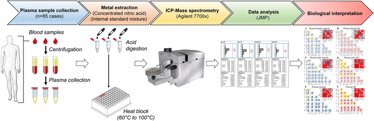
Materials and methods

Ethics

We performed this case–control study under a protocol approved by the Leeds Teaching Hospitals NHS Trust Research Ethics Committee. All procedures accorded with the ethics committee guidelines with informed consent from all participants or families of patients as appropriate.

Patient selection

Patients and controls without cognitive impairment were respectively recruited (Vardy et al. 2009) through memory clinics in Leeds and Dewsbury (England) and the Leeds Family Health Services Authority day hospitals and elderly medicine outpatient clinics in the Leeds area. All were of European Caucasian background and gave written informed consent (consent from relatives of the AD-patients was provided, where appropriate). Diagnosis of probable AD was made in accordance with international diagnostic criteria (National Institute of Neurological and Communicative Disorders and Stroke–Alzheimer’s Disease and Related Disorders Association Work Group: NINCDS-ADRDA) (McKhann et al. 1984). All participants underwent a standardised clinical evaluation: medical history, fasting plasma glucose (FPG) and HbA1c (a measure of chronic glycaemia), and cognitive function assessment by Mini-Mental State Examination (MMSE). AD samples were selected by excluding patients with diagnosed T1D or T2D (including those taking insulin). Samples were then selected from the whole-study population for whom required measurements (FPG, HbA1c) were available (Vardy et al. 2007). The resulting 42 AD-patients were then age- and gender-matched to 43 controls (Table 1).

Table 1

Characteristics of participants in this study
Characteristic
AD
Control
P value
Number
42
43
Male (%)
52.4
53.5
NS
Age (years)
78.2 ± 1.3
78.1 ± 1.1
NS
MMSE score
21 (11–25)
29 (27–30)
< 0.001
ApoE4 allele-positive (%)
71.4
32.6
< 0.001
Fasting plasma glucose (mmol/l)
5.0 (3.8–10.6)
5.1 (4.2–8.9)
NS
Serum HbA1ca
5.6 (5.1–7.8)
5.7 (5.1–8.4)
NS
a Hemoglobin A1c

Sample digestion

Plasma samples of 50 μL were digested in 2-mL microcentrifuge tubes (Eppendorf) as described below. Detailed method descriptions are available (Xu et al. 2017). Reactions were performed in concentrated nitric acid (A509 Trace Metal Grade; Fisher, Loughborough, UK) to which had been added 5% (v/v) Agilent Internal Standard mixture (5183-4681; Agilent Technologies, Cheadle, UK). This internally standardised acid was also used at appropriate dilutions to provide rinse and calibration solutions, at 2% (v/v) final nitric acid concentration.
Calibration solutions were produced by appropriate dilutions of Environmental Calibration Standard (Agilent 5183-4688). Acid digestion was carried out using an ‘open-vessel’ method. The tube lids were punctured to prevent pressure build up, and 0.2 ml standard-containing nitric acid added. Tubes were then inserted into a “Dri-block” heater, which was initially at room temperature. In parallel, 50-µl aliquots of NIST SRM 1950 plasma (Standard Reference Material; National Institute of Standards and Technology, Gaithersburg, MD), were processed in the same way. Tubes with standard-containing acid but no sample were also processed in each batch to provide “digestion” blanks. Temperature was then set to 60 °C and the block switched on. After 30 min, the set temperature was increased to 100 °C, and digestion continued for a further 210 min. After digestion, the tubes were allowed to cool overnight.
Aliquots of 100 μL were taken from each digestion solution and added to 15-mL Falcon tubes (525–0629, VWR, Lutterworth, UK) containing 5 mL LC–MS grade water, to produce solutions for analysis at a final nitric acid concentration of 2% (v/v).

Metal measurements

Tissue metal concentrations were measured using an Agilent 7700× ICP-MS spectrometer equipped with a MicroMist nebulizer (Glass Expansion, Melbourne, Australia) and a Scott double-pass spray chamber. Sample introduction was performed using an Agilent Integrated autosampler (I-AS) with helium as the collision gas. A multi-element method including all elements present in the calibration solution was applied, as previously reported by our group (Church et al. 2015; Xu et al. 2017). Calibration solutions were produced by appropriate dilutions of Environmental Calibration Standard (Agilent 5183-4688). An intermediate concentration from this calibration series was used throughout as a periodic quality control sample. Instrument and digestion blanks were also interspersed throughout the experiment and the detection limit for each element determined by comparison of calibration samples and blanks. Scandium was used as the internal standard for all elements except Zn and Se, where germanium was used. Two collision-cell gas modes were applied: all elements were analysed in helium mode (5.0 ml/min helium), except for Se which was analysed in high-energy helium mode (10 ml/min helium). Germanium internal standard was analysed in both modes. Mode selection followed Agilent recommendations to minimise interference for measured elements by e.g. isobaric cluster ions. Integration times were: 0.1 s for Na, Mg, K and Ca; 0.3 s for Cu and Zn; 0.01 s for Fe; and 3 s for Se. NIST SRM plasma samples were also analysed within this experiment to check for precision of our measurements when compared to the certified values.

Statistical methods

The analysis for case–control study group characteristics was performed using SPSS Statistics 22 (IBM). Distributions of variables were tested for skewness using the Kolmogorov–Smirnov Goodness-Of-Fit test, for 42 AD participants and 43 matched controls (Table 1). Parametric data are presented as mean ± standard error of the mean (SEM), and non-parametric data as median (range). Comparisons between AD and control groups were performed using an Independent-Samples t test for parametric, and the Mann–Whitney U test for non-parametric data. Pearson’s χ2 analysis was performed on categorical data.
ICP-MS data were exported to Microsoft Excel and log-transformed for statistical analysis. Means (± 95% CI) of the log-transformed data were calculated and the significance of between-group differences was examined by unpaired t-tests with Welch’s correction to allow for unequal variances and sample sizes. Means (± 95% CI) were back-transformed to reflect the actual concentrations of elements. Statistical calculations were performed using GraphPad v6.04 (Prism; La Jolla, CA). P values of < 0.05 have been considered significant, and those of 0.05 ≤ P < 0.10 have also been tabulated. For correlation analyses, serial pairwise correlations were generated between the reported metal abundances (log10 transformed) using the statistical software JMP® (Version 12.0.0; SAS Institute, Cary, NC). The correlations generated were visually depicted using scatter plot matrices and those considered as “strong” (Pearson coefficient R > 0.5 (or < − 0.5) and P values < 0.001) were further investigated. The statistical significance of differences in correlation coefficients between strong correlations in controls and AD groups was determined by Fisher r-to-z transformation and resulting P values < 0.05 (two-tailed) were considered significant.

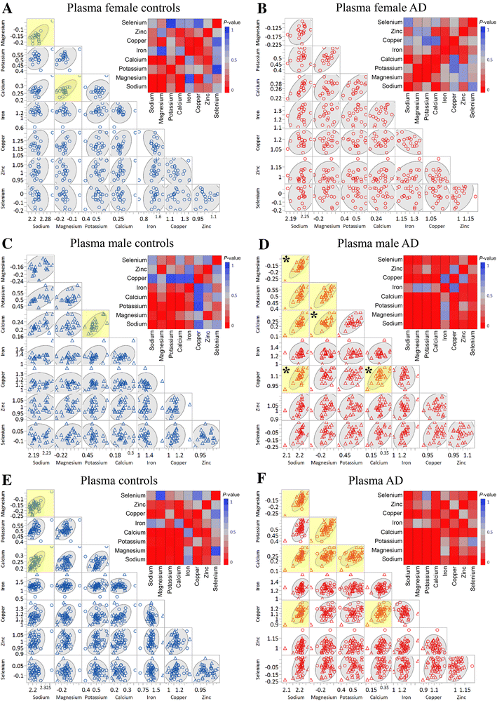
Results and discussion

In this study, we measured concentrations of seven essential metals and the metalloid, Se, in plasma samples from 42 cases with AD and 43 matched control participants (Table 1). We matched participants in this case–control study for gender and age (Table 1). Consistent with expectation, measured cognitive function (MMSE scores) was significantly lower in the AD group than controls whereas the ApoE4 allele was more prevalent. Levels of FPG and serum HbA1c were equivalent between groups. There was no evidence for elevated rates of undiagnosed T2D or impaired glucose tolerance (IGT)/impaired fasting glucose (IFG) in this group of British patients with sporadic AD (Table 1). Therefore, the current study compares plasma metal levels in AD cases representative of sporadic AD with appropriately matched controls.
We rigorously controlled the application of the ICP-MS methodology and have a high degree of confidence in the robustness of our data. For example, we included NIST SRM plasma samples in our study as previously described (Xu et al. 2017) and verified that measurements were within the ± 5% difference range compared to the certified values, indicating the precision of the methodology applied in this study.
We analysed the female and male datasets separately, and then combined them, in order to highlight possible effects of gender in control and AD participants (Table 2, Fig. 1). In the female patients, none of the eight measured elements differed in their plasma concentrations between the AD and control groups (P = 0.021; Table 2). However, in male patients, there was modest evidence that Zn trended towards increase in the AD group compared to the control group (Table 2). This might be consistent with previously-reported elevations in levels of Zn in AD brain (Schrag et al. 2011), and possibly also with enrichment of Zn in and around amyloid plaques (Lovell et al. 1998). However, reports concerning elevations of Zn in AD brain are not uniform. For example, in a recent metallomic study, brain-Zn levels in AD were moderately elevated in only three of seven regions studied: entorhinal cortex, middle temporal gyrus, and cerebellum (Xu et al. 2017). By contrast, they were similar to control values in other regions known to contain high levels of amyloid plaques in AD. Thus, the relationship between bulk regional Zn concentrations and Zn in amyloid plaques is not entirely clear. Since multi-regional studies of brain metal content are sparse, it is difficult to reach firm conclusions concerning the occurrence or significance of elevated brain-Zn levels in AD.

Table 2

Plasma metal concentrations in groups of females, males and combined sexes
Element (reference isotope)
Concentration unit
Groups
Control
AD
Pvalue
Na (23Na)
mmol/L
F
170 (166–175)
167 (164–169)
NS
M
160 (158–162)
162 (156–167)
NS
F & M
165 (162–167)
164 (161–167)
NS
Mg (24Mg)
mmol/L
F
0.70 (0.67–0.73)
0.70 (0.69–0.74)
NS
M
0.70 (0.66–0.71)
0.70 (0.68–0.73)
NS
F & M
0.70 (0.67–0.71)
0.70 (0.69–0.73)
NS
K (39K)
mmol/L
F
3.3 (3.2–3.5)
3.2 (3.0–3.4)
NS
M
3.3 (3.1–3.4)
3.3 (3.1–3.4)
NS
F & M
3.3 (3.2–3.4)
3.3 (3.1–3.4)
NS
Ca (44Ca)
mmol/L
F
1.9 (1.8–1.9)
1.8 (1.8–1.9)
NS
M
1.7 (1.6–1.7)
1.8 (1.7–1.8)
0.087
F & M
1.8 (1.7–1.8)
1.8 (1.7–1.8)
NS
Fe (56Fe)
μmol/L
F
17.6 (14.5–21.3)
17.8 (16.3–19.5)
NS
M
20.1 (16.9–24.1)
18.4 (16.7–20.4)
NS
F & M
18.9 (16.6–21.5)
18.1 (17.0- 19.4)
NS
Cu (63Cu)
μmol/L
F
15.9 (14.6–17.3)
14.5 (13.3–15.8)
NS
M
13.2 (12.1–14.3)
12.7 (11.7–13.9)
NS
F & M
14.4 (13.5–15.3)
13.5 (12.7–14.4)
NS
Zn (66Zn)
μmol/L
F
10.2 (9.8–10.7)
10.5 (9.5–11.6)
NS
M
10.0 (9.6–10.4)
10.8 (10.2–11.5)
0.021*
F & M
10.1 (9.8–10.4)
10.7 (10.1–11.2)
0.075
Se (78Se)
μmol/L
F
0.90 (0.84–0.98)
1.00 (0.89–1.05)
NS
M
0.90 (0.86–1.01)
0.90 (0.81–0.95)
NS
F & M
0.90 (0.87–0.97)
0.90 (0.86–0.97)
NS
Data are means (± 95% CI); P values for significance of between-group differences were calculated by applying Welch’s modified t tests to metal measurements from plasma samples from female patients (F) [control (n = 20) and AD (n = 19)], male patients (M) [control (n = 23) and AD (n = 23)], and all patients combined (F & M) [control (n = 43) and AD (n = 42)]
*Significant (P values < 0.05)

Fig. 1

Correlations between metal levels in the plasma of AD cases and controls. Scatter plots of metal abundance correlations are shown for each metal measured in control (blue) and AD (red) plasma in females (ab), male (cd) and combined sexes (ef). Shown are Pvalues corresponding to each metal-pair, with correlations as a gradient colour in the heat map for each gender group. All the strong correlations (R > 0.5 (or < − 0.5) and Pvalues of < 0.001) are highlighted in yellow and, for each correlation, grey ellipsoids represent the 95% confidence interval. Note that the metal co-regulations were only perturbed in the male AD cases whereas values were unaffected in females or combined groupings. *Significantly different correlations (Fisher r-to-z transformation P values < 0.05) between control and AD participants in the male group. Legend: control males (blue triangles); control females (blue circles); AD males (blue circles); AD females (red circles). (Color figure online)
In addition, the observation of elevated serum-Zn levels in males but not females with AD is an intriguing finding. Some workers have suggested that altered Zn homeostasis could play a significant role in AD pathogenesis, since it can clearly interact with APP and Aβ, and reportedly participates in APP processing, and aggregation and clearance of Aβ in the AD-brain. Data strongly support the enrichment of Zn in the core of Aβ plaques, a process that has been suggested to disrupt Zn homeostasis in brain regions important for memory generation and vulnerable to AD pathogenesis (Craddock et al. 2012; Miller et al. 2006). However, we note that interventions aimed at ameliorating alterations in Zn homeostasis have no accepted role in the management of AD at the time of writing.
In our combined analyses that included separate analysis of data from both genders, none of the measured metals showed statistically significant between-group concentration differences (Table 2). Therefore, our results suggest that alterations in plasma metal levels in association with AD may be inconsistent between genders. In line with our observations, a previous study concluded that metal levels are not consistently altered in plasma of AD participants, except for an elevated abundance of mercury (Gerhardsson et al. 2008). Other studies have reported decreased plasma levels of Mg, Cu, Zn, Fe and Se (Vural et al. 2010) and serum levels of Zn (Huang et al. 2013) in patients with AD compared with controls using different approaches, but the findings of our current study are not consistent with these prior reports, perhaps due to the minor differences of method employed. However, we would note that the evidence generated from our study is strongly supported by the methods employed and the use of the NIST SRM as a control to verify the efficacy of the digestion and metal recovery and measurements processes [for further data concerning the performance of our methods, see (Xu et al. 2017)].
We also performed an exploratory pairwise correlational analysis to ascertain how the measured metals might be differently co-regulated between controls and cases. Firstly, the number of strong correlations (R > 0.5 (or < − 0.5) and P values < 0.001) was determined for each group (Table 3, Fig. 1). We observed a higher number of strong correlations in the all-AD group (~ 18% increase; Table 3), a finding possibly consistent with a systemic alteration of metal co-regulation in these patients. Notably, when we divided the groups by gender, only the AD-male group showed an increase in strong correlations (~ 21.5% increase). In addition, in the male group there were significant changes between pairwise correlations in metal levels between the controls and cases. Significant differences included changes in the following metal–metal pairs: magnesium-sodium (Rcontrol = 0.41, RAD = 0.84; P = 0.02); calcium-magnesium (Rcontrol = 0.28, RAD = 0.73; P = 0.04); copper-sodium (Rcontrol = − 0.03, RAD = 0.65; P = 0.009); and copper-calcium (Rcontrol = − 0.02, RAD = 0.65; P = 0.01). These copper-associated correlations were the most significant differences observed in AD-male participants when compared to controls (Fig. 1a–f). Interestingly, in another study in patients with AD, substantively elevated sodium levels were present in all three severely damaged brain regions assessed: hippocampus, entorhinal cortex, and middle temporal gyrus. Copper was markedly decreased in these regions as well, but also in four other brain regions studied (Xu et al. 2017). Therefore, there is some evidence for a copper-sodium interaction in the brain of patients with AD, which may be related to the finding of a sodium-copper correlation in the plasma.

Table 3

Number of correlations for each group
Gender groupa
Strong correlations controls
Strong correlations AD
Shared correlations controls/AD
Female (F)
2 (7.1%)
0 (0%)
0 (0%)
Male (M)
1 (3.6%)
7 (25.0%)
0 (0%)
Combined (F & M)
2 (7.1%)
7 (25.0%)
2 (7.1%)
aIn each group, 28 correlations were tested for significance
T2D has strong epidemiological ties with AD, and T2D is known to cause significant perturbations in the systemic homeostasis of numerous essential metals (Cooper et al. 2005). It was therefore important to eliminate it from both groups in the current study, to exclude any possible confounding effect from participants with known T2D or those with undiagnosed disease. We did this by history, and screening all potential participants for T2D using FPG and HbA1c measurements. This is a precaution that should be undertaken in future studies of systemic metal regulation in AD, in order to avoid a possible confounding effect by untoward inclusion of patients with T2D, particularly given reports of increased prevalence of T2D in populations of patients with AD (Janson et al. 2004). In the current study therefore, alterations in metal homeostasis detected in participants with AD were not due to possible T2D.
While plasma levels of individual metals are of limited informational value here, how these metals are differentially co-regulated may represent a more useful measurement for further investigation as potential diagnostic biomarker of AD status. Likewise, other groups have indicated correlation coefficient matrix-assessed correlations as a suitable method to enable biomarker discovery in CNS-associated disorders (Nahan et al. 2017). Indeed, correlation analyses may assist in unmasking global dysregulation of systemic metal homeostasis in AD in a more comprehensive way as many of the pathways where metals play distinctive roles are mutually interconnected. As just one example, Cu plays a major role in the regulation of systemic uptake and distribution of Fe (Nittis and Gitlin 2002). Therefore, we suggest that larger scale studies are needed to verify the alteration of plasma metal levels in AD and we indicate the monitoring of co-regulated plasma metals as a relevant measurement to be tested as potential diagnostic biomarker in AD.
Finally, our results have significance for guiding future investigations on the utility of plasma metals as potential peripheral biomarkers to track rapidly progression or severity in subjects with AD, and possibly to predict disease onset before clinical and neurological symptoms are clearly manifested. In addition, plasma metals might be relevant as pharmacodynamic/pharmacokinetic biomarkers to facilitate clinical studies involving metal chelation treatment in AD.

Notes

Acknowledgements

We thank Cynthia Tse, University of Auckland, for reviewing the manuscript. This study was funded by The University of Manchester, the Central Manchester University Hospitals NHS Foundation Trust, the Northwest Regional Development Agency, by project grants from the Medical Research Council (MR/L010445/1) and Biotechnology and Biological Sciences Research Council (BB/I018654/1), and was facilitated by the Manchester Biomedical Research Centre and the Greater Manchester Comprehensive Local Research Network. GJSC also receives research funding and support from Endocore Research Associates (60187; 3601607); the Maurice and Phyllis Paykel Trust; Lottery Health (New Zealand); the Health Research Council (New Zealand); and a programme grant from the Ministry for Business, Innovation, and Employment (New Zealand; UOAX9902; PMIX0201; UOAX0705; UOAX0815). All data are provided in full in the results section of this manuscript.

Compliance with ethical standards

Conflict of interest

GJSC is named as inventor on patents disclosing Cu-selective chelators for the treatment of the diabetic complications and related diseases: these patents have been licensed and he has no financial interest in this work and declares no other duality of interest. All the other authors declare that they have no duality of interest with respect to this manuscript.

References

  1. Adlard PA, Bush AI (2006) Metals and Alzheimer’s disease. J Alzheimer’s Dis 10:145–163CrossRefGoogle Scholar
  2. Akatsu H et al (2012) Transition metal abnormalities in progressive dementias. Biometals 25:337–350.  https://doi-org.ezp.welch.jhmi.edu/10.1007/s10534-011-9504-8CrossRefPubMedGoogle Scholar
  3. Ayton S, Lei P, Bush AI (2013) Metallostasis in Alzheimer’s disease. Free Radic. Biol. Med 62:76–89.  https://doi-org.ezp.welch.jhmi.edu/10.1016/j.freeradbiomed.2012.10.558CrossRefPubMedGoogle Scholar
  4. Bush AI (2013) The metal theory of Alzheimer’s disease. J Alzheimer’s Dis 33:S277–281.  https://doi-org.ezp.welch.jhmi.edu/10.3233/jad-2012-129011Google Scholar
  5. Church SJ et al (2015) Deficient copper concentrations in dried-defatted hepatic tissue from ob/ob mice: a potential model for study of defective copper regulation in metabolic liver disease. Biochem Biophys Res Commun 460:549–554.  https://doi-org.ezp.welch.jhmi.edu/10.1016/j.bbrc.2015.03.067CrossRefPubMedPubMedCentralGoogle Scholar
  6. Citron M (2010) Alzheimer’s disease: strategies for disease modification. Nat Rev Drug Discov 9:387–398.  https://doi-org.ezp.welch.jhmi.edu/10.1038/nrd2896CrossRefPubMedGoogle Scholar
  7. Cooper GJS et al (2005) Demonstration of a hyperglycemia-driven pathogenic abnormality of copper homeostasis in diabetes and its reversibility by selective chelation. Diabetes 54:1468–1476.  https://doi-org.ezp.welch.jhmi.edu/10.2337/diabetes.54.5.1468CrossRefPubMedGoogle Scholar
  8. Craddock TJ, Tuszynski JA, Chopra D, Casey N, Goldstein LE, Hameroff SR, Tanzi RE (2012) The zinc dyshomeostasis hypothesis of Alzheimer’s disease. PLoS ONE 7:e33552.  https://doi-org.ezp.welch.jhmi.edu/10.1371/journal.pone.0033552CrossRefPubMedPubMedCentralGoogle Scholar
  9. Ferri CP et al (2005) Global prevalence of dementia: a Delphi consensus study. Lancet 366:2112–2117CrossRefPubMedPubMedCentralGoogle Scholar
  10. Fiandaca MS, Mapstone ME, Cheema AK, Federoff HJ (2014) The critical need for defining preclinical biomarkers in Alzheimer’s disease. Alzheimer’s Dement 10:S196–212.  https://doi-org.ezp.welch.jhmi.edu/10.1016/j.jalz.2014.04.015CrossRefGoogle Scholar
  11. Gerhardsson L, Lundh T, Minthon L, Londos E (2008) Metal concentrations in plasma and cerebrospinal fluid in patients with Alzheimer’s disease. Dement Geriatr Cogn Disord 25:508–515.  https://doi-org.ezp.welch.jhmi.edu/10.1159/000129365CrossRefPubMedGoogle Scholar
  12. Huang X et al (1999) The A beta peptide of Alzheimer’s disease directly produces hydrogen peroxide through metal ion reduction. Biochemistry 38:7609–7616.  https://doi-org.ezp.welch.jhmi.edu/10.1021/bi990438fCrossRefPubMedGoogle Scholar
  13. Huang X, Moir RD, Tanzi RE, Bush AI, Rogers JT (2004) Redox-active metals, oxidative stress, and Alzheimer’s disease pathology. Ann NY Acad Sci 1012:153–163CrossRefPubMedGoogle Scholar
  14. Huang CW, Wang SJ, Wu SJ, Yang CC, Huang MW, Lin CH, Cheng IH (2013) Potential blood biomarker for disease severity in the Taiwanese population with Alzheimer’s disease. Am J Alzheimer’s Dis Other Dement 28:75–83.  https://doi-org.ezp.welch.jhmi.edu/10.1177/1533317512467674CrossRefGoogle Scholar
  15. Janson J, Laedtke T, Parisi JE, O’Brien P, Petersen RC, Butler PC (2004) Increased risk of type 2 diabetes in Alzheimer disease. Diabetes 53:474–481.  https://doi-org.ezp.welch.jhmi.edu/10.2337/diabetes.53.2.474CrossRefPubMedGoogle Scholar
  16. Kaddurah-Daouk R et al. (2013) Alterations in metabolic pathways and networks in Alzheimer/’s disease, Transl Psychiatry 3:e244. http://www.nature.com.ezp.welch.jhmi.edu/tp/journal/v3/n4/suppinfo/tp201318s1.html
  17. Lovell MA, Robertson JD, Teesdale WJ, Campbell JL, Markesbery WR (1998) Copper, iron and zinc in Alzheimer’s disease senile plaques. J Neurol Sci 158:47–52CrossRefPubMedGoogle Scholar
  18. McKhann G, Drachman D, Folstein M, Katzman R, Price D, Stadlan EM (1984) Clinical diagnosis of Alzheimer’s disease: report of the NINCDS-ADRDA Work Group under the auspices of department of health and human services task force on Alzheimer’s disease. Neurology 34:939–944CrossRefPubMedGoogle Scholar
  19. Miller LM, Wang Q, Telivala TP, Smith RJ, Lanzirotti A, Miklossy J (2006) Synchrotron-based infrared and X-ray imaging shows focalized accumulation of Cu and Zn co-localized with beta-amyloid deposits in Alzheimer’s disease. J Struct Biol 155:30–37.  https://doi-org.ezp.welch.jhmi.edu/10.1016/j.jsb.2005.09.004CrossRefPubMedGoogle Scholar
  20. Nahan KS, Walsh KB, Adeoye O, Landero-Figueroa JA (2017) The metal and metalloprotein profile of human plasma as biomarkers for stroke diagnosis. J Trace Elem Med Biol 42:81–91.  https://doi-org.ezp.welch.jhmi.edu/10.1016/j.jtemb.2017.04.004CrossRefPubMedGoogle Scholar
  21. Nittis T, Gitlin JD (2002) The copper-iron connection: hereditary aceruloplasminemia. Semin Hematol 39:282–289CrossRefPubMedGoogle Scholar
  22. Prince M, Guerchet M, Prina M (2013) Policy brief for heads of government: the global impact of dementia 2013–2050. Alzheimer’s Dis Int 2013:1–8Google Scholar
  23. Ramos P, Santos A, Pinto NR, Mendes R, Magalhaes T, Almeida A (2014) Anatomical region differences and age-related changes in copper, zinc, and manganese levels in the human brain. Biol Trace Elem Res 161:190–201.  https://doi-org.ezp.welch.jhmi.edu/10.1007/s12011-014-0093-6CrossRefPubMedGoogle Scholar
  24. Roberts BR, Ryan TM, Bush AI, Masters CL, Duce JA (2012) The role of metallobiology and amyloid-beta peptides in Alzheimer’s disease. J Neurochem 120:149–166.  https://doi-org.ezp.welch.jhmi.edu/10.1111/j.1471-4159.2011.07500.xCrossRefPubMedGoogle Scholar
  25. Sastre M, Ritchie CW, Hajji N (2015) Metal ions in alzheimer’s disease brain. JSM Alzheimer’s Dis Relat Dement 2:1014Google Scholar
  26. Schrag M et al (2011) Effect of cerebral amyloid angiopathy on brain iron, copper, and zinc in Alzheimer’s disease. J Alzheimer’s Dis 24:137–149.  https://doi-org.ezp.welch.jhmi.edu/10.3233/jad-2010-101503Google Scholar
  27. Smith DG, Cappai R, Barnham KJ (2007) The redox chemistry of the Alzheimer’s disease amyloid beta peptide. Biochem Biophys Acta 1768:1976–1990.  https://doi-org.ezp.welch.jhmi.edu/10.1016/j.bbamem.2007.02.002CrossRefPubMedGoogle Scholar
  28. Squitti R (2012) Metals in alzheimer’s disease: a systemic perspective. Front Biosci 17(1):451CrossRefGoogle Scholar
  29. Vardy ER, Rice PJ, Bowie PC, Holmes JD, Grant PJ, Hooper NM (2007) Increased circulating insulin-like growth factor-1 in late-onset Alzheimer’s disease. J Alzheimers Dis 12:285–290
———–

https://www.health.harvard.edu/blog/zinc-for-the-common-cold-not-for-me-201102171498

Zinc for the common cold? Not for me


Former Executive Editor, Harvard Health
Media channels are atwitter with the news that zinc can beat the common cold. CBS News, the L.A. Times, the Huffington Post, and hundreds of others are treating a quiet research report as big news that will have a life-changing effect. After reading the report and doing a little digging into the dark side of zinc, I’m not rushing out to stock up on zinc lozenges or syrup.
The latest hubbub about zinc was sparked by a report from the Cochrane Collaboration. This global network of scientists, patients, and others evaluates the evidence on hundreds of different treatments. In the latest review, on zinc for the common cold, researchers Meenu Singh and Rashmi R. Das pooled the results of 13 studies that tested zinc for treating colds. By their analysis, taking zinc within 24 hours of first noticing the signs of a cold could shorten the cold by one day. They also found that taking zinc made colds a bit less severe.
Sounds good so far. But instead of saying, “Hey, take zinc if you have a cold,” the researchers concluded like this: “People taking zinc lozenges (not syrup or tablet form) are more likely to experience adverse events, including bad taste and nausea. As there are no studies in participants in whom common cold symptoms might be troublesome (for example, those with underlying chronic illness, immunodeficiency, asthma, etc.), the use of zinc currently cannot be recommended for them. Given the variability in the populations studied (no studies from low- or middle-income countries), dose, formulation and duration of zinc used in the included studies, more research is needed to address these variabilities and determine the optimal duration of treatment as well as the dosage and formulations of zinc that will produce clinical benefits without increasing adverse effects [italics mine], before making a general recommendation for zinc in treatment of the common cold.”
Not exactly a ringing endorsement.
Zinc isn’t something to mess around with. Two years ago, the FDA warned everyone to stop using zinc-containing nasal sprays to fight colds because these sprays had been linked to more than 100 cases of loss of smell. The Institute of Medicine set the tolerable upper limit for zinc at 40 milligrams a day for adults, less for teens and children. The tolerable upper limit is the highest daily intake “likely to pose no risk of adverse health effects for almost all individuals.” If you follow the directions on zinc-based cold remedies, you’ll get more than the tolerable upper limit.
The glowing media reports are sure to spark the next cold rush as supplement makers crank up their zinc output. I’m going to ignore it. I don’t like the taste of zinc lozenges, and don’t think it’s worth sucking on them for several day to knock a day off a cold.
At the first sign of a cold, I’m headed to the kitchen to make chicken noodle soup.

Other Articles waiting to access:

 2018;62(3):1319-1335. doi: 10.3233/JAD-170732.

A Long Journey into Aging, Brain Aging, and Alzheimer’s Disease Following the Oxidative Stress Tracks.

Abstract

The Editors of the Journal of Alzheimer’s Disease invited Professor Patrizia Mecocci to contribute a review article focused on the importance and implications of her research on aging, brain aging, and senile dementias over the last years. This invitation was based on an assessment that she was one of the journal’s top authors and a strong supporter of the concept that oxidative stress is a major contributor to several alterations observed in age-related conditions (sarcopenia, osteoporosis) and, more significantly, in brain aging suggesting a pivotal role in the pathogenesis and progression of one of the most dramatic age-related diseases, Alzheimer’s disease (AD). Her first pioneering research was on the discovery of high level of 8-hydroxy-2′-deoxyguanosine (OH8dG), a marker of oxidation in nucleic acids, in mitochondrial DNA isolated from cerebral cortex. This molecule increases progressively with aging and more in AD brain, supporting the hypothesis that oxidative stress, a condition of unbalance between the production of reactive oxygen species and antioxidants, gives a strong contribution to the high incidence of AD in old age subjects. OH8dG also increases in peripheral lymphocyte from AD subjects, suggesting that AD is not only a cerebral but also a systemic disease. The role of antioxidants, particularly vitamin E and zinc, were also studied in longevity and in cognitive decline and dementia. This review shows the main findings from Mecocci’s laboratory related to oxidative stress in aging, brain aging, and AD and discusses the importance and implications of some of the major achievements in this field of research.

 2018;62(3):1369-1379. doi: 10.3233/JAD-170662.

Metals and Alzheimer’s Disease: How Far Have We Come in the Clinic?

Abstract

It is estimated that by the year 2050 there will be more than 1.5 billion people globally over the age of 65 years. Aging is associated with changes to a number of different cellular processes which are driven by a variety of factors that contribute to the characteristic decline in function that is seen across multiple physiological domains/tissues in the elderly (including the brain). Importantly, aging is also the primary risk factor for the development of neurodegenerative disorders such as Alzheimer’s disease. As such, there is an urgent need to provide a greater understanding of both the pathogenesis and treatment of these devastating neurodegenerative disorders. One of the key cellular processes that becomes dysregulated with age and participates both directly and indirectly in age-related dysfunction, is metal homeostasis and the neurochemistry of metalloproteins, the basic science of which has been extensively reviewed in the past. In this review, we will focus on the human clinical intervention trials that have been conducted over approximately the last four decades that have attempted to establish the efficacy of targeting metal ions in the treatment of AD.
Shopping Cart