Conjunctivochalasis:
Conjunctivochalasis (CCH) is a common condition in which there are extra folds of the conjunctiva, the clear covering over the sclera (the white part of the eye). CCH can cause chronic eye pain, irritation, foreign body sensation, redness, tearing, burning. If conservative treatments do not help (lid hygiene with warm compresses, artificial tears, increased Omega 3 intake, Doxycycline pills, Xiidra, Restasis, Lipiflow, Intense Pulse Light for the Meibomian glands if one has Meibomian Gland Dysfunction, etc), we recommend the following options to treat the CCH and get rid of your complaints.
1. Argon Laser: this laser is applied to the conjunctiva, through it, and into the sclear surface. This laser tries to tack back down the conjunctiva to the sclera.
2. Surgical Excision: If the Argon Laser does not help, then we excise the extra, redundant conjunctiva with a surgical excision. There are many ways this can be done:
a. Surgical excision of extra conjunctival tissue with placement with amniotic membrane (obtained in a sterile condition and tested for safety from a placenta of a newborn baby from FDA approved companies who have obtained these placentas from mothers who have given their consent).
More information below from these great sites:
1.https://www.eyeworld.org/article-conjunctivochalasis
OCTOBER 2011
|
CORNEA
|
Conjunctivochalasis: An overlookedbut commonailment
by Michelle Dalton EyeWorld Contributing Editor |
|||
|
Treatment varies, but surgery is usually necessary

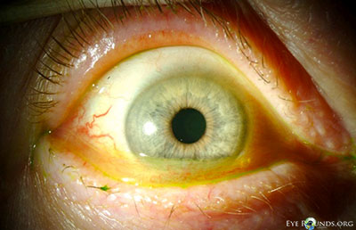
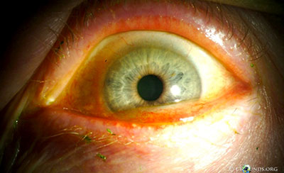
The excess conjunctival folds (stained) indicative of conjunctivochalasis Source: Steven G. Safran, M.D.
 Conjunctivochalasis is easier to identify after Lissamine green staining Source: Steven G. Safran, M.D. Conjunctivochalasis is easier to identify after Lissamine green staining Source: Steven G. Safran, M.D.
Although not uncommon, conjunctivochalasis may be overlooked or mistaken for an age-related change comprising redundant conjunctival folds between lower lid margins and the globe. “Many times I have sat in the room listening to patients complain of vague on again/off again foreign body sensation, typically after cataract surgery, and have been unable to give an answer for their problems other than dry eye. However, some of these patients presented without the typical corneal signs of dryness and had adequate tear film stability,” said Gary Wortz, M.D., private practice, Bluegrass Eye Surgery, Lebanon, Ky. “This subset of patients bothered me, and through further research and careful examination, I have been able to identify conjunctivochalasis as a common yet often overlooked culprit.” When there is refractory conjunctivochalasis, surgical resection with or without amniotic graft transplantation is typical. The issue with chalasis “is that there is a deficiency of Tenon’s capsule, and thus the conjunctiva is not just redundant but does not have the proper adherence to the underlying tissue,” said Steven G. Safran, M.D., private practice, Lawrenceville, N.J. He has “been treating conjunctival chalasis surgically for many years. My current approach is to use the Ellman (Oceanside, N.Y.) radio surgical unit with a Teflon-tipped white 0.04-mm probe. This technique has been published,1 and I’ve been using it since. It shrinks the conjunctiva in a series of applications under/through the conjunctiva, and it’s fast, easy, and reasonably effective. If that fails, I do resection with an amniotic membrane graft that works very effectively but in a few cases can lead to fibrosis/redness that persists for months and may not be cosmetically acceptable. It’s also far more expensive and bothersome to take patients to the OR for this than to fix it in the office.”
Dr. Safran said resecting and in-office suturing used to be his preferred treatment strategy, but he abandoned that technique when persistent chemosis “took months to resolve” in rare instances. In Dr. Safran’s experience, radio-wave electrosurgery addresses both the deficiency and the adherence issue. “In severe cases, resection will still be required, and amniotic grafts work well in these cases most of the time,” he said. In his amniotic graft technique, surgery is performed under topical anesthesia with lidocaine gel and “straight epinephrine drizzled on the eye for hemostasis,” he said. He noted surgeons should expect “very little bleeding” when they cut into the loose conjunctivochalasis tissue. Also, moisten the cornea “every now and then,” but not to the point where it might disturb the amniotic graft or the tissue glue. Although he’s performing more of these types of treatments, Dr. Safran maintains his “go-to” surgery is radiosurgery. He is now performing a “more aggressive procedure” on cases with a lot of excess tissue.
“I’ve been grabbing the excess tissue with a clamp [a modified foldable IOL forceps] and using the Ellman with a pointed cautery tip to ‘melt away’ the excess conjunctiva while the clamp creates a new ‘seal’ at the base,” Dr. Safran said. “I can get a greater reduction of excess tissue in moderate to severe cases with this technique without resorting to surgical removal in the OR. This is a modified version of the technique described by Stephen Pflugfelder, M.D.2 In his technique he uses cautery and calls it ‘thermoreduction.’ In my technique I use the Ellman and find it works better and with less inflammation and more control than cautery.”
A “great pearl” for minimizing bleeding during conjunctival surgery is to “drop topical epinephrine on the ocular surface before dissection. There is almost no bleeding except from the limbal vessels. It was astonishing to me when I first did it because I didn’t think it had enough time to react, but it works great,” said D. Brian Kim, M.D., in private practice, Professional Eye Associates, Dalton, Ga. He said he learned this technique from Scheffer C.G. Tseng, M.D., director, Ocular Surface Center, Miami, Fla.; medical director, Ocular Surface Research & Education Foundation (OSREF), Miami, Fla.; and director of research & development, Tissue Tech, Miami, Fla. Videos and discussions of the topic are available on OSREF’s website: www.osref.org/conjunctivochalasis-cch.aspx.
At the 2010 World Cornea
Congress and American Academy of Ophthalmology conference, Linden Reed Doss, R. Philip Doss, M.D., and E. Lauren Doss described a new surgical technique for the treatment of conjunctivochalasis in which the redundant conjunctiva is precisely resected. The AAO video (“Paste-Pinch-Cut: A novel surgical repair for conjunctivochalasis”) was awarded Best of Show in the Cornea, External Disease topic. Basically, the technique involved creating an arc-like guideline that is demarcated inferior to the limbus. A small buttonhole is made in the temporal bulbar conjunctiva at the edge of the marking line. Fibrin glue is injected through the buttonhole along the line. The conjunctiva is pinched with modified (curved) ptosis forceps gathering the excess conjunctiva into a ridge at the top of which lies the marking line. The authors noted the forcep curve follows the globe line. This ridge is excised after 20 seconds (the amount of time needed for the glue to coagulate), leaving a sealed wound. The authors also said that in 139 eyes of 70 patients, conjunctivochalasis resolved in all patients, and 91.5% reported improvement in symptoms. More work is being done to understand the underlying causes of conjunctivochalasis. A recent study3 analyzed protein profiles in the tears of healthy patients and those with conjunctivochalasis and found 24 spots were “significantly upregulated in conjunctivochalasis compared with that in controls,” the authors wrote, adding that some of the proteins are markers of inflammation and oxidative processes.
References
1. Youm DJ, Kim JM, Choi CY. Simple Surgical Approach with High-Frequency Radio-Wave Electrosurgery for Conjunctivochalasis. Ophthalmology. 2010;117:21292133.
2. Gumus K, Crockett CH, Pflugfelder SC. Anterior segment optical coherence tomography: a diagnostic instrument for conjunctivochalasis. Am J Ophthalmol. 2010;150(6):798-806.
3. Acera A, Suarez T, Rodriguez-Agirretxe I, Vecino E, Duran JA. Changes in tear protein profile in patients with conjunctivochalasis. Cornea. 2011;30(1):42-9.
|
||||
February 6, 2013
Chief Complaint: Watery, irritated eyes
History of Present Illness: This patient was a healthy 76-year-old woman with complaints of irritated, red, and watery eyes. She had been followed in our clinic for these concerns and was being managed with doxycycline 100 mg daily, Lotemax® (loteprednol etabonate ophthalmic suspension) daily to both eyes, artificial tears, and artificial tear ointment to both eyes. She felt this regimen had helped relieve some of her symptoms, but was still bothered by ocular irritation, intermittent epiphora, and blurriness of her vision while reading.
Examination:
Visual Acuity with Correction:
- Right eye (OD): 20/20
- Left eye (OS): 20/20-3
Intraocular Pressure (mm Hg)
- OD: 14
- OS: 15
Slitlamp Examination:
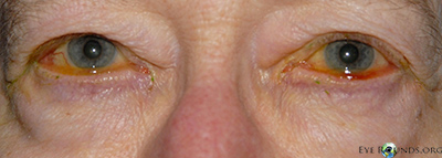
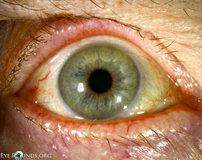
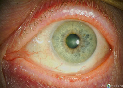
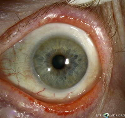
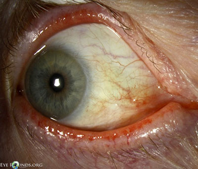
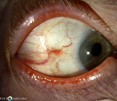
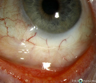
- External examination: Conjunctivochalasis apparent on gross examination
- Lids: dermatochalasis, meibomian gland dysfunction, no lagophthalmos, both eyes (OU)
- Anterior segment: marked conjunctivochalasis, corneas clear without punctate epithelial erosions, anterior chamber deep and quiet, normal iris architecture, 1-2+ nuclear sclerosis
- Posterior segment: within normal limits OU
 |
 |
 |
 |
 |
Given her symptoms and failure to respond to medical management as well as the marked conjunctivochalasis, the patient elected to proceed with surgery. The operative plan included resection of the excessive inferior and nasal bulbar conjunctiva with placement of an amniotic membrane graft. She underwent the procedure first on her right eye and noted a great amount of improvement in her symptoms, especially the blurriness while reading. The same operative procedure was performed four months later in her left eye. She was followed for over one year after surgery on her right eye (and over six months postoperatively on her left eye) and continued to be symptomatically improved compared to preoperatively.
Video: Conjunctival resection with placement of AMT (amniotic membrane transplant) Conjunctival resection with amniotic membrane graft for conjunctivochalasis
If video fails to load, see http://vimeo.com/59018451
 |
 |
 |
 |
 |
 |
Discussion:
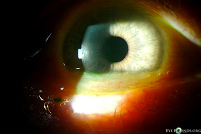
Conjunctivochalasis (CCh) is defined as redundant conjunctiva. Hughes first coined this entity in 1942; [1] however the description of loose, nonedematous conjunctiva had been first reported as early as 1908 by Elschnig.[2] It is most often evident between the globe and the lower eyelid, but in more advanced cases can be evident around the entire globe. The majority of cases are bilateral, and often conjunctivochalasis is overlooked as a normal variant associated with the aging process if the patient is asymptomatic. In cases where the patient is symptomatic, common symptoms include: tearing, foreign body sensation, ocular irritation, and blurriness, especially in down gaze. It is important to keep this condition in the differential of chronic ocular irritation and epiphora.
Conjunctivochalasis is a common finding among older adults. Studies suggest that conjunctivochalasis is more common in patients who have dry eye and meibomian gland disease/blepharitis and is associated with contact lens wear.[3] The etiology of conjunctivochalasis is not well understood. Theories include that it could be a natural aging process of the conjunctiva or that it could be due to lid position abnormalities, ocular movements, ocular irritation, and eye rubbing. Some histopathologic studies demonstrate elastosis, chronic, nongranulomatous inflammation, fragmentation of the elastic fibers, and loss of collagen. Matrix metalloproteinases (MMPs) are enzymes that modify or degrade the extracellular matrix. MMP-1 and MMP-3 enzymes have been noted to be overexpressed in conjunctivochalasis fibroblasts in tissue culture, while the enzyme levels of tissue inhibitors of metalloproteinases (TIMPs) are unchanged. The change in the balance between these two groups of enzymes may facilitate the breakdown of the extracellular changes and lead to the clinically evident changes observed in conjunctivochalasis.[4] Another hypothesis is that pressure from the eyelids may lead to impaired lymphatic drainage of the conjunctiva, which is supported by findings of lymphangiectasia, fragmentation of the elastic fibers, and no signs of inflammation on histopathology.[5]
If the patient has conjunctivochalasis but is asymptomatic, no treatment is necessary. For symptomatic patients, medical treatment is recommended as the initial step. Medical management includes the use of ocular lubricants, antihistamines (if there is a component of allergic conjunctivitis), and topical steroids. In cases where medical management is insufficient in improving the patient’s symptoms, surgical intervention may be necessary. Surgical management is directed at resecting the redundant conjunctival tissue. Several methods have been described in the literature. The most common methods of surgical intervention include a crescent-shaped conjunctival resection with or without sutures, resection with placement of an amniotic membrane graft (with either sutures or fibrin tissue glue or both), or suture fixation of the redundant conjunctiva to the globe (without resection). During procedures where the conjunctiva is resected, a crescent of tissue can be marked, excised, and left to heal or closed with absorbable suture. Another method of resection is to make a limbal peritomy, extend posteriorly with relaxing incisions, and then pull the conjunctiva anteriorly, resecting the excess tissue that extends past the limbus.[6] The conjunctiva is then re-approximated near the limbus. Lastly, amniotic membrane grafting is an option. Once the area of conjunctiva is excised, an amniotic membrane graft is secured to the globe corresponding to the excised conjunctival defect, secured with fibrin tissue glue or absorbable suture, or a combination of the two.[7,8]
Success rates of the various methods appear to be similar. Moderate to high rates of improvement in symptoms have been reported. In a study by Yokoi and colleagues, an improvement of symptoms was found in 88.2% of patients that underwent resection of symptomatic conjunctivochalasis.[6] Similar success rates were reported by Tseng and colleagues.[8]
| Grade | Number of folds and relationship to the tear meniscus height |
|---|---|
| 1 | No persistent fold |
| 2 | Single, small fold |
| 3 | More than two folds and not higher than the tear meniscus |
| 4 | Multiple folds and higher than the tear meniscus |
Diagnosis: conjunctivochalasis
|
|
|
|
Differential Diagnosis:
- Chemosis
- Conjunctival infection or inflammation
- Entropion
- Meibomian gland disease
References:
- Hughes WL. Conjunctivochalasis. Am J Ophthalmology 1942;25:48-51.
- Elschnig A. Beitrag zur Aethiologie und Therapie der cronischen Konjunctivitis. Dtsch Med Wochenschr 1908; 26: 1133-1155.
- Mimura T, Yamagami S, Usui T, et al. Changes of conjunctivochalasis with age in a hospital-based study. Am J Ophthalmology. 2009;147:171-177.
- Li DQ, Meller D, Liu Y, Tseng SC. Overexpression of MMP-1 and MMP-3 by cultured conjunctivochalasis fibroblasts. Invest Ophthalmol Vis Sci. 2000;41:404-410.
- Wattanabe A, Yokoi N, Kinoshita S, et al. Clinicopathologic study of conjunctivochalasis. Cornea. 2004;23:294-298.
- Yokoi N, Komuro A, Nishii M, et al. Clinical impact of conjunctivochalasis on the ocular surface. Cornea. 2005;24:524-531.
- Serrano F, Mora LM. Conjunctivochalasis: a surgical technique. Ophthalmic Surgery. 1989;20:883-884.
- Meller D, Maskin SL, Pires RT, Tseng SC. Amniotic membrane transplantation for symptomatic conjunctivochalasis refractory to medical treatments. Cornea. 2000;19:796-803.
- Meller D, Tseng SC. Conjunctivochalasis: literature review and possible pathophysiology. Survey of Ophthalmology. 1998; 43: 225-232.