
When Lipiflow first came out in 2011, many eyeMDs and ODs were very skeptical. Would it really do anything? Would it help patient relieve their dry eye symptoms? Would it help regenerate meibomian glands? I too was very skeptical. At an initial price of $2000 when it first came out, it seemed to expensive for most patients to even try it.
Years later, we are seeing many patients improve after Lipiflow. While it is technically indicated to save meibomian glands, I am seeing many patients feel a significant improvement and even being pain free after Lipiflow. It is not a guarantee, but it is worth having performed if you have enough glands remaining. If you have too few glands left, it might be too late to have Lipiflow, which does not hurt and usually takes about 12 minutes.
I am seeing more and more patients have a clear improvement on meibography after the Lipiflow. A previous post is noted below, but the below photos are of a patient who feel almost immediate relief of chronic eye burning and her meibomian gland structure clearly improved.
https://drcremers.com/2017/11/another-patient-who-felt-significant.html
This patient below’s symptoms went from a 8 out 10 pain to about a 2 out of 10 pain.
The oil that is expressed at her lid margin with lid pressure also looks more clear and less thick.
Look at the infrared images also. You can see that her right lower lid had what appears to be absent-glands and they seemed to have filled up. 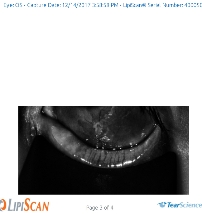


This is the photo of the Right Eye Before Lipiflow (11/2/2017) and After 12/14/17.
You can see improvement in the filling of the meibomian gland both on the regular photo and on the infrared photo. No PRP was injected.


This is the left eye Before Lipiflow (11/2/2017) and After 12/14/17.
The left eye also looks a bit better.

This is the Infrared photo of the left eye Before Lipiflow (11/2/2017) and After 12/14/17. You can see less “black” empty spaces after the Lipiflow.

Soon there will be no one in this country or world who fits “a” as everyone is on some sort of screen more than 4hr a day it seems…..especially kids.