Dry Eye Syndrome and Hormones

Dry eyes can be very frustrating in older women, especially if they feel their hormone replacement therapy is making the dry eye worse.

Here is a great summary of what to be aware of with hormone levels in general and dry eyes.

SLC

Hormones and Dry Eye Syndrome

An Update on What We Do and Don’t Know

Eduardo Melani Roch, Flavio Mantelli, Luis Fernando Nominato, Stefano Bonini

Disclosures

Curr Opin Ophthalmol. 2013;24(4):348-355. 

Abstract

Purpose of review Dry eye syndrome (DES) prevalence is large and its relationship with hormonal diseases is becoming clearer, although more complex. This review provides insight to this association as well as clarifying what remains unanswered about how to interpret and treat findings common to both DES and hormonal diseases.
Recent findings Several sex hormone-related diseases are associated with DES. Hormone replacement therapy to correct such conditions has conflicting outcomes based on epidemiologic studies and clinical trials. Thyroid-associated diseases are frequently involved in DES and must be investigated in cases where the cause of the ocular disease is undetermined. Diabetes mellitus is one of the major causes of DES, whereas correcting the metabolic imbalance minimizes its ocular symptomology. Gene therapy to treat DES-related hormonal diseases is a promising option based on animal studies.
Summary Diagnosis and management of hormonal diseases can minimize the ocular surface damage and severity of DES. Clinical care of DES includes patient evaluation of hormonal status. Future research requires clarification of the underlying disease mechanisms and identifying novel strategies to reprogram the endocrine system rather than chronic medication usage.

Introduction

See which places are the best and worst to practice medicine, and why.


As early as 1930, Dr Henrik Sjögren indicated that there is an association between hormonal changes and dry eye syndrome (DES).[1] His first study mentioned the disease only in women and had one patient with glucose tolerance test results reflective of an insulin resistance. Since then, numerous other results confirmed an association between endocrine system balance, ocular surface homeostasis and lacrimal gland function.[2] Moreover, the role of hormonal diseases in DES progression is now more extensively described providing novel perspectives for hormonal treatment of this condition.
Currently, it is apparent that in several different diseases caused by a hormonal imbalance, DES clinical findings occur (Table 1).
This review analyzes recent controversy surrounding the relationship among menopause changes and DES. It focuses also on the association between DES and the most common endocrine diseases involving changes in sex hormones, thyroid-associated diseases, and diabetes mellitus. A complementary topic describes the potential use of gene therapy to reprogram lacrimal gland function so as to overcome hormonal disease-induced ocular surface damage.

Sex Hormones

Dry eye syndrome prevalence is much higher among women and aging is a risk factor for this condition. This realization suggests that sex hormones are key factors on this condition and output during menopause could worsen DES, whereas sex hormone replacement therapy (SHRT) may instead attenuate this condition. However, epidemiologic studies are not supportive of this idea as DES incidence in women on SHRT is even greater than that in individuals not undergoing such treatment.[9] Specifically, they showed that there is instead a higher incidence of DES among older women on SHRT, especially those using estrogen alone. As a matter of fact, with longer-term SHRT use, DES frequency and symptomology increased.[9] These findings disagree with other studies in which menopause was found to be a risk factor for DES, but SHRT was instead of some benefit in those cases.[10,11] On the contrary, another epidemiologic study confirmed that SHRT is a DES risk factor [odds ratio (OR) 1.6, 95% confidence interval (CI) 1.0–2.5].[12] This study agrees with other observations indicating that estrogen therapy in women triggered or worsened the onset of DES and/or Sjögren’s syndrome.[13–15]
A possible explanation for these conflicting conclusions is that the outcome of SHRT depends on estrogen dosage; age of the individuals when therapy is first initiated, namely, estrogen may be only beneficial in younger persons, whereas detrimental and/or pro-inflammatory in postmenopausal women; and type and combination of SHRT applied. It has been demonstrated that estrogen administered at physiological doses is supportive of lacrimal gland function and preservation of anterior ocular surface health at early ages, but at higher doses and/or in combination with other hormonal supplements would be injurious and/or induce inflammation. Elderly women would be more susceptible to these aforementioned limitations of SHRT. This expectation was confirmed by a study showing that higher estrogen levels correlated with declines in tear secretion in women older than 60 years, whereas in women who were in their fourth decade tear output instead improved.[16,17]
Moreover, some studies showed that DES symptoms identified with the ocular surface disease index (OSDI) questionnaire worsened with SHRT using drospirenone and estradiol, but Schirmer test and tear film break-up time (TFBUT) improved after 6 months of treatment. This dichotomy suggests that it is difficult to arrive at a meaningful conclusion regarding the safety and or efficacy of DES treatment as it is a multifactorial disease. Furthermore, any comparative study based on only one or a few parameters monitored for just a limited time is inadequate for deciding how to manage this disease.[14,18]
Women with Sjögren’s syndrome are deficient in certain types of androgen such as dihydrotestosterone and dehydroepiandrosterone (DHEA), but present with similar levels of testosterone and estrogen compared to age-matched controls.[19] Systemic treatment of Sjögren’s syndrome patients with DHEA only partially reverse its lower saliva levels, despite marked increase in the blood level of DHEA.
Endometriosis is a gynecological disease whose pathophysiology involves higher estrogen levels. There are recent conflicting reports addressing the association of Sjögren’s syndrome with this disease and therefore with estrogen.[20,21]
See which places are the best and worst to practice medicine, and why.


Such differences support the notion of the uncertainty of the SHRT in treating DES ( Table 2 ). SHRT is problematic because sex hormone action is affected by endocrine production levels, circulating hormonal levels of total and free forms, peripheral tissue hormonal conversion, target cell receptor and enzymatic affinity, interactions with effects of other stimuli, and final physiologic action of thousands of genes regulated by sex hormones in tissues responsible for tear film and ocular surface health maintenance. Those aforementioned factors are also known as intracellular or intracrine hormonal phenomena in peripheral tissues. Their involvement is not completely understood and may also account for variable findings in regard to the association between changes in sex hormonal levels and DES.[22-24]
Therefore, therapeutic testing on an individual basis is necessary in menopausal women to verify whether or not SHRT is beneficial in treating DES. Moreover, to confirm sex hormone dysfunctional involvement in DES, a clinical gynecologic investigation maybe complemented by measuring the following sex hormone serum levels: total testosterone, free testosterone, 17-hydroxiprogesterone, dihydroepiandrosterone sulfate (SDHEA), in the early follicular phase of the menstrual cycle. In addition, thyroid function tests as described below and prolactin levels are also useful to eliminate other endocrine diseases. Those tests can be complemented by ultrasonographic ovary analysis.

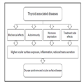
Thyroid Hormone-related Diseases and Dry Eye Syndrome
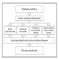
Ocular surface and tear film changes are very common in patients with thyroid disease.[25–29] It is also clinically relevant to consider occult thyroid disease in the differential diagnosis of DES patients.[30]
There is some ambiguity in the medical literature pertaining to identifying the association between thyroid disease and involvement of DES. Sometimes the terminology does not clarify the exact disease and in other instances an assignment is not definitive because groups of different diseases are lumped together as a single entity ( Table 3 ).
Although DES in thyroid disorders is usually considered as complication of an autoimmune condition related to Hashimoto’s thyroiditis and/or Graves’ ophthalmopathy, there are several causes of a thyroid disorder with diverse underlying mechanisms probably contributing to induce DES in those patients.
In these diseases, a combination of events contributes to manifesting DES in the patients. The most obvious one is an ocular surface disturbance due to enhanced environmental exposure and lid mechanical impairment in Graves’ ophthalmopathy. Inappropriate lid closure is caused by superior eyelid retraction, eye globe proptosis, and impaired blinking. All these factors are contributory to inadequate tear film surface spreading and higher evaporation (Fig. 1).[31-33]

Figure 1.


Aspect of the lid opening (a) and ocular surface (b) of a patient with Graves’ ophthalmopathy.
In recent years, the concomitance between thyroid diseases and Sjögren’s syndrome and more detailed descriptions of ocular surface inflammatory responses in patients with thyroid diseases made it likely that DES in those patients is an autoimmune-induced response.[26,34,35] Although there are no known reports on lacrimal gland pathology in DES individuals afflicted with a thyroid disease, computed tomography analysis revealed that those glands were larger than controls.[36] The biopsies of salivary glands revealed infiltrating lymphocytes (mainly CD3+ T) with a CD4+/CD8+ ratio of 2 : 1, activation markers, human leukocyte antigen (HLA) class II molecules, and interleukin (IL)-2 receptor (CD25). In some patients, HLA class II was inappropriately expressed in the epithelial gland cells.[26]
Autoantibodies against thyroid stimulating hormone (TSH) receptor are expressed in individuals with thyroid-associated ophthalmopathy and these TSH receptors are present in human lacrimal gland (LG).[37] In addition, autoantibodies against thyroid hormone were observed in thyroid diseases such as Hashimoto’s thyroiditis and Graves’ disease, but also in Sjögren’s syndrome. These observations suggest a mechanism whereby autoimmunity disrupts hormone and tissue interaction, due to antibody binding to hormones and/or their cognate receptors, leading to LG dysfunction.[38]
Impaired thyroid hormone activity, per se, can be responsible for LG impairment and DES, since in rats LG express thyroid hormone receptor beta-1. Furthermore, the antithyroid drug thiamazole, given systemically to rats, induced tear secretion reduction and cornea epithelial metaplasia.[39] A clinical study revealed also that thyroid hormone regulates lipid exocrine secretion, which suggests its impact on meibomian glands and evaporative DES.[40]
Get an inside look at what happens when physicians get sued for malpractice.


The thyroid hormone disorder treatments include thyroid hormone replacement, iodine suppression, immunomodulators specific for the orbit and ocular disease, local radiotherapy, and oculoplastic and orbit surgical interventions.[26,41,42] It is difficult to identify in the present medical literature whether each one of them contribute to DES development and/or progression. This uncertainty exists since clinical trials targeted to address these two questions are not available and the data are only from case reports. In a study involving a follow-up for 9 years of patients receiving various treatments for Graves’ ophthalmopathy, about 25% of them presented with DES complaints.[43] In another study, testing surgical millerectomy for eyelid retraction caused by Graves’ ophthalmopathy, DES, identified by Schirmer test measurement, was observed in 7 of the 12 patients. In response to orbit radiotherapy, also for Graves’ ophthalmopathy, persistent DES symptoms were reported in 10 and 12% of patients.[44,45]
Taken together, these findings reveal that various factors present in the spectrum and long-term progression of ocular surface findings associated to thyroid diseases. Some of the contributory factors to DES include increased exposure, inflammatory responses, and hormonal level reduction (Fig. 2).

Figure 2.


Mechanisms of disease related to DES in thyroid diseases. DES, dry eye syndrome.
These considerations indicate that in every DES case lacking a clear cause, these individuals should also be evaluated for thyroid disease. Such screening entails a clinical exam and serum analysis for thyroxin (T4) and TSH. Moreover, in these cases, apart from ocular topical treatment, the patients may benefit from endocrine treatment, but the ophthalmologist needs to be aware of possible ocular side effects resulting from Graves’ ophthalmopathy treatment.

Diabetic Dry Eye

Dry eye syndrome is common in diabetic patients, which makes diabetes mellitus one of the most frequent among the known DES causes.[46–49]
Diabetes mellitus is caused by insulin insufficiency or peripheral tissue resistance to this hormone.[50] Since insulin is a ubiquitous hormone responsible for many events at the cellular level and diabetes mellitus causes complications in various organs and systems that interact with the LG and ocular surface, it is difficult to point to a single underlying mechanism responsible for diabetic DES. Possible mechanisms include decline in insulin hormone expression, LG and ocular surface neuropathy, vascular impairment, and systemic hyperosmotic disturbance (Fig. 3).[51–54]

Figure 3.


Mechanisms of disease related to DES in diabetes mellitus.
In diabetic patients, the frequency of DES varies from 15.4 to 82%. Independently whether they are type I or type II, adult or children, the most frequent reported signs and symptoms are lower TFBUT and tear secretion measured by Schirmer test and conjunctival metaplasia.[55–57] DES may be exacerbated by chronic hyperglycemia, diabetes mellitus duration, and local triggers such as ocular surface trauma or intraocular surgery. This disease seems to be more severe in patients with advanced stages of diabetic retinopathy.[53,54] However, it is not clear whether the severity is just a consequence of the uncontrolled metabolic status and/or side effects of systemic drugs. Other possibilities include multiple surgical manipulation and other interventions to restore or preserve some vision.
Among the aforementioned studies, there are variations among the type and number of parameters used to identify DES in diabetes mellitus. Therefore, better tests are still needed to detect alone diabetic DES, as well as the relationship between ocular and clinical systemic parameters. The reason for this uncertainty is that the patients included in the various studies are heterogeneous in terms of age, disease duration, and clinical control.
Adequate insulin levels are necessary for LG, and ocular surface maintenance and function since in culture this hormone is crucial for acinar LG cell and cornea epithelial cell proliferation.[58,59] Moreover, tear insulin levels are reflective of its systemic levels. Its systemic deprivation in diabetic animal models induces LG size reduction and several histological and molecular changes that are partially reversed by systemic insulin application.[60–62] A clinical study showed that hypoinsulinemia is an independent risk factor for polyneuropathy.[63] These findings may suggest that insulin deprivation or resistance may cause nerve-conduction abnormalities and reduced secretion in association with corneal pain.
Topical insulin application was proposed to treat corneal epithelial defects, not just those related to diabetes mellitus. Its positive effect was observed in rabbits and rats.[46,64–66]
Which specialists are sued most often?


The systemic osmotic imbalance of diabetes mellitus may translate into tear film instability and DES symptoms as recently observed in a clinical study, not specifically addressing diabetes mellitus.[67] In diabetic patients, tear film instability and higher osmolarity may be explained by higher glucose and protein levels in tears and changes in the protein profile.[68] These changes may be influenced by the time length and glucose level control of the diabetes mellitus condition, and also offer a rationale for a diagnostic tool to monitor plasma glycemic levels based on tear glucose levels.[68,69] A recent intent to apply the tear glycemic analysis was unsuccessful.[68]
Diabetic peripheral neuropathic pain is present in 5–42% of individuals with diabetes mellitus. There is a possibility that at some point in the course of this disease (diabetes mellitus) the manifestation of neuropathy in the ocular surface contributes to signs and symptoms of DES.[63,70]
Clinical and experimental evidence indicates that diabetes mellitus causes of DES are multifactorial. The high frequency of diabetes mellitus in the population and DES in diabetic patients indicates that it is relevant to test for diabetes mellitus in DES patients in case of any clinical suspicion. This entails a combined effort among clinicians to periodically determine if these patients’ metabolic parameters and ocular health are being controlled. Although the clinical presentation of DES in diabetes mellitus is very heterogeneous and can be already present at an early stage of diabetes mellitus, clinical studies reveal that poorly controlled diabetes mellitus increases the frequency and DES severity in the long term.[53,54]

Gene Therapy for LG

Various hormones are essential contributors for maintaining LG function. Such control sustains tear film composition and volume needed to nourish and protect the anterior ocular surface.[2] Under several different conditions involving hormonal expression dysregulation, medications can induce hormonal replacement needed to restore the hormonal balance, required to inhibit or suppress immune responses.
In situations where only chronic and/or local hormonal doses can meet these needs, gene therapy, however, may be a viable alternative. Such an approach has been used in different animal models to deliver a gene of interest with a viral vector.[71-73] The possibility of using noncritical-for-life organs like salivary and lacrimal glands has been shown to be well tolerated and efficient for local and systemic therapeutic purposes in these models.[74]
What emotional and financial effects do physicians experience from a malpractice lawsuit?


Growth hormones, along with erythropoietin expanding-4 (a promoter of insulin secretion), were transfected into salivary glands. Insulin expression was detected in saliva or blood of mice.[75,76] In animal models of dry mouth and dry eye induced by radiotherapy, the treated group presented with reduced frequency of oral ulcers, improved cornea epithelial thickness, and restoration of the production of saliva and tears.[77]
The concept of applying virus vector carried gene therapy to treat ocular surface diseases, using the lacrimal gland as an endogenous bioreactor, was recently evaluated. The tropism of certain serotypes of adeno-associated virus vector for specific target lacrimal gland cells, and the expression of the luciferase protein without histological or functional damage was described.[78]
These findings suggest that the lacrimal gland is a feasible target for gene therapy in cases of DES related to hormonal dysfunction. Current indications are that hormonal production could be induced in this peripheral organ without compromising local or systemic health. Such therapy also has the potential to be of benefit to other local or regional tissues.

Conclusion

We describe here the current knowledge base pertaining to the role of some hormonal imbalances in DES initiation and progression. Clinical findings and pathophysiological mechanisms confirm the associations between diabetes mellitus, estrogen level declines, and thyroid gland diseases and DES. Very promising preliminary results obtained in several animal models are described for employing gene therapy as a novel alternative to improve treatment of hormone related DES.

References

  1. Murube J. Henrik Sjögren, 1899–1986. Ocul Surf 2010; 8:2–7.
  2. Sullivan DA, Wickham LA, Rocha EM, et al. Influence of gender, sex steroid hormones, and the hypothalamic-pituitary axis on the structure and function of the lacrimal gland. Adv Exp Med Biol 1998; 438:11–42.
  3. Mantelli F, Moretti C, Micera A, Bonini S. Conjunctival mucin deficiency in complete androgen insensitivity syndrome (CAIS). Graefes Arch Clin Exp Ophthalmol 2007; 245:899–902.
  4. Sullivan BD, Evans JE, Cermak JM, et al. Complete androgen insensitivity syndrome: effect on human meibomian gland secretions. Arch Ophthalmol 2002; 120:1689–1699.
  5. Krenzer KL, Dana MR, Ullman MD, et al. Effect of androgen deficiency on the human meibomian gland and ocular surface. J Clin Endocrinol Metab 2000; 85:4874–4882.
  6. Coksuer H, Ozcura F, Oghan F, et al. Effects of hyperandrogenism on tear function and tear drainage in patients with polycystic ovary syndrome. J Reprod Med 2011; 56:65–70.
  7. Bonini S, Mantelli F, Moretti C, et al. Itchy-dry eye associated with polycystic ovary syndrome. Am J Ophthalmol 2007; 143:763–771.
  8. Alves M, Dias AC, Rocha EM. Dry eye in childhood: epidemiological and clinical aspects. Ocul Surf 2008; 6:44–51.
  9. Schaumberg DA, Buring JE, Sullivan DA, Dana MR. Hormone replacement therapy and dry eye syndrome. J Am Med Assoc 2001; 286:2114–2119.
  10. Jensen AA, Higginbotham EJ, Guzinski GM, et al. A survey of ocular complaints in postmenopausal women. J Assoc Acad Minor Phys 2000; 11:44–49.
  11. Okon A, Jurowski P, Gos R. The influence of the hormonal replacement therapy on conjunctival epithelium morphology among peri- and postmenopausal women. Klinika oczna 2001; 103:183–186.
  12. Chia EM, Mitchell P, Rochtchina E, et al. Prevalence and associations of dry eye syndrome in an older population: the Blue Mountains Eye Study. Clin Exp Ophthalmol 2003; 31:229–232.
  13. Nagler RM, Pollack S. Sjögren’s syndrome induced by estrogen therapy. Semin Arthritis Rheum 2000; 30:209–214.
  14. Erdem U, Ozdegirmenci O, Sobaci E, et al. Dry eye in postmenopausal women using hormone replacement therapy. Maturitas 2007; 56:257–262.
  15. Uncu G, Avci R, Uncu Y, et al. The effects of different hormone replacement therapy regimens on tear function, intraocular pressure and lens opacity. Gynecol Endocrinol 2006; 22:501–505.
  16. Mathers WD, Stovall D, Lane JA, et al. Menopause and tear function: the influence of prolactin and sex hormones on human tear production. Cornea 1998; 17:353–358.
  17. Wenderlein M, Mattes S. The ‘dry eye’ phenomenon and ovarian function. Study of 700 women pre and postmenopausal. Zentralblatt fur Gynakologie 1996; 118:643–649.
  18. Coksuer H, Ozcura F, Oghan F, et al. Effects of estradiol-drospirenone on ocular and nasal functions in postmenopausal women. Climacteric 2011; 14:482–487.
  19. Sullivan DA, Belanger A, Cermak JM, et al. Are women with Sjögren’s syndrome androgen-deficient? J Rheumatol 2003; 30:2413–2419.
  20. Haga HJ, Gjesdal CG, Irgens LM, Ostensen M. Reproduction and gynaecological manifestations in women with primary Sjögren’s syndrome: a case-control study. Scand J Rheumatol 2005; 34:45–48.
  21. Nielsen NM, Jorgensen KT, Pedersen BV, et al. The co-occurrence of endometriosis with multiple sclerosis, systemic lupus erythematosus and Sjögren syndrome. Hum Reprod 2011; 26:1555–1559.
  22. Sullivan DA, Jensen RV, Suzuki T, Richards SM. Do sex steroids exert sex-specific and/or opposite effects on gene expression in lacrimal and meibomian glands? Molec Vis 2009; 15:1553–1572.
  23. Porola P, Straub RH, Virkki LM, et al. Failure of oral DHEA treatment to increase local salivary androgen outputs of female patients with Sjögren’s syndrome. Scand J Rheumatol 2011; 40:387–390.
  24. Labrie F, Labrie C. DHEA and intracrinology at menopause, a positive choice for evolution of the human species. Climacteric 2013; 16:205–213.

    * This review explains the intracrine events related to DHEA in postmenopausal women, suggesting that it is a positive evolutionary process.

  25. Galor A, Feuer W, Lee DJ, et al. Prevalence and risk factors of dry eye syndrome in a United States veterans affairs population. Am J Ophthalmol 2011; 152:377–384.e2.
  26. Coll J, Anglada J, Tomas S, et al. High prevalence of subclinical Sjögren’s syndrome features in patients with autoimmune thyroid disease. J Rheumatol 1997; 24:1719–1724.
  27. Mergler S, Garreis F, Sahlmuller M, et al. Thermosensitive transient receptor potential channels in human corneal epithelial cells. J Cell Physiol 2011; 226:1828–1842.
  28. Pan Z, Wang Z, Yang H, et al. TRPV1 activation is required for hypertonicity-stimulated inflammatory cytokine release in human corneal epithelial cells. Invest Ophthalmol Vis Sci 2011; 52:485–493.
  29. Yang H, Wang Z, Capo-Aponte JE, et al. Epidermal growth factor receptor transactivation by the cannabinoid receptor (CB1) and transient receptor potential vanilloid 1 (TRPV1) induces differential responses in corneal epithelial cells. Exp Eye Res 2010; 91:462–471.
  30. Pan Z, Yang H, Reinach PS. Transient receptor potential (TRP) gene superfamily encoding cation channels. Hum Genomics 2011; 5:108–116.
  31. Garcia DM, Barbosa JC, Pinto CT, Cruz AA. Estimation of spontaneous blinking main sequence in normal subjects and patients with Graves’ upper eyelid retraction. Invest Ophthalmol Vis Sci 2013; 54:1434–1442.

    ** This study compares the characteristics of blinking in Graves’ and control individuals.

  32. Brasil MV, Brasil OF, Vieira RP, et al. Tear film analysis and its relation with palpebral fissure height and exophthalmos in Graves’ ophthalmopathy. Arq Bras Oftalmol 2005; 68:615–618.
  33. Gilbard JP, Farris RL. Ocular surface drying and tear film osmolarity in thyroid eye disease. Acta Ophthalmol (Copenh) 1983; 61:108–116.
  34. Biro E, Szekanecz Z, Czirjak L, et al. Association of systemic and thyroid autoimmune diseases. Clin Rheumatol 2006; 25:240–245.
  35. Jara LJ, Navarro C, Brito-Zeron Mdel P, et al. Thyroid disease in Sjögren’s syndrome. Clin Rheumatol 2007; 26:1601–1606.
  36. Harris MA, Realini T, Hogg JP, Sivak-Callcott JA. CT dimensions of the lacrimal gland in Graves orbitopathy. Ophthal Plast Reconstructr Surg 2012; 28:69–72.

    * This study establishes correlation between the lacrimal gland morphology and a clinical disease using a noninvasive method.

  37. Eckstein AK, Finkenrath A, Heiligenhaus A, et al. Dry eye syndrome in thyroid-associated ophthalmopathy: lacrimal expression of TSH receptor suggests involvement of TSHR-specific autoantibodies. Acta Ophthalmol Scand 2004; 82 (3 Pt 1):291–297.
  38. Ruggeri RM, Galletti M, Mandolfino MG, et al. Thyroid hormone autoantibodies in primary Sjögren syndrome and rheumatoid arthritis are more prevalent than in autoimmune thyroid disease, becoming progressively more frequent in these diseases. J Endocrinol Invest 2002; 25:447–454.
  39. Dias AC, Modulo CM, Jorge AG, et al. Influence of thyroid hormone on thyroid hormone receptor beta-1 expression and lacrimal gland and ocular surface morphology. Invest Ophthalmol Vis Sci 2007; 48:3038–3042.
  40. Goolamali SK, Evered D, Shuster S. Thyroid disease and sebaceous function. Br Med J 1976; 1:432–433.
  41. Gurdal C, Genc I, Sarac O, et al. Topical cyclosporine in thyroid orbitopathy-related dry eye: clinical findings, conjunctival epithelial apoptosis, and MMP-9 expression. Curr Eye Res 2010; 35:771–777.
  42. Bartley GB, Fatourechi V, Kadrmas EF, et al. The treatment of Graves’ ophthalmopathy in an incidence cohort. Am J Ophthalmol 1996; 121:200–206.
  43. Bartley GB, Fatourechi V, Kadrmas EF, et al. Long-term follow-up of Graves ophthalmopathy in an incidence cohort. Ophthalmology 1996; 103:958–962.
  44. Prabhu RS, Liebman L, Wojno T, et al. Clinical outcomes of radiotherapy as initial local therapy for Graves’ ophthalmopathy and predictors of the need for postradiotherapy decompressive surgery. Radiat Oncol 2012; 7:95–102.
  45. Matthiesen C, Thompson JS, Thompson D, et al. The efficacy of radiation therapy in the treatment of Graves’ orbitopathy. Int J Radiat Oncol Biol Phys 2012; 82:117–123.

    * The study followed more than 200 patients along of 10 years and presented a rate of success of 84% of Graves’ ophthalmopathy treatment with radiotherapy. However, it also shows that 12% of patients developed DES.

  46. Wang TJ, Wang IJ, Hu CC, Lin HC. Comorbidities of dry eye disease: a nationwide population-based study. Acta Ophthalmol 2012; 90:663–668.

    ** This is a national epidemiologic study conducted in Taiwan with more than 10 000 individuals. It identified the comorbidities of DES.

  47. Amos AF, McCarty DJ, Zimmet P. The rising global burden of diabetes and its complications: estimates and projections to the year 2010. Diabet Med 1997; 14 (Suppl 5):S1–S85.
  48. King H, Aubert RE, Herman WH. Global burden of diabetes, 1995–2025: prevalence, numerical estimates, and projections. Diabetes Care 1998; 21:1414–1431.
  49. Kaiserman I, Kaiserman N, Nakar S, Vinker S. Dry eye in diabetic patients. Am J Ophthalmol 2005; 139:498–503.
  50. Saad MJ. Molecular mechanisms of insulin resistance. Braz J Med Biol Res 1994; 27:941–957.
  51. Rocha EM, de MLMH, Carvalho CR, et al. Characterization of the insulin-signaling pathway in lacrimal and salivary glands of rats. Curr Eye Res 2000; 21:833–842.
  52. Azar DT, Spurr-Michaud SJ, Tisdale AS, Gipson IK. Altered epithelial-basement membrane interactions in diabetic corneas. Arch Ophthalmol 1992; 110:537–540.
  53. Dogru M, Katakami C, Inoue M. Tear function and ocular surface changes in noninsulin-dependent diabetes mellitus. Ophthalmology 2001; 108:586–592.
  54. Nepp J, Abela C, Polzer I, et al. Is there a correlation between the severity of diabetic retinopathy and keratoconjunctivitis sicca? Cornea 2000; 19:487–491.
  55. Akinci A, Cetinkaya E, Aycan Z. Dry eye syndrome in diabetic children. Eur J Ophthalmol 2007; 17:873–878.
  56. Goebbels M. Tear secretion and tear film function in insulin dependent diabetics. Br J Ophthalmol 2000; 84:19–21.
  57. Rahman A, Yahya K, Ahmed T, Sharif-Ul-Hasan K. Diagnostic value of tear films tests in type 2 diabetes. J Pak Med Assoc 2007; 57:577–581.
  58. Hann LE, Kelleher RS, Sullivan DA. Influence of culture conditions on the androgen control of secretory component production by acinar cells from the rat lacrimal gland. Invest Ophthalmol Vis Sci 1991; 32:2610–2621.
  59. Lindberg K, Brown ME, Chaves HV, et al. In vitro propagation of human ocular surface epithelial cells for transplantation. Invest Ophthalmol Vis Sci 1993; 34:2672–2679.
  60. Cunha DA, Carneiro EM, Alves Mde C, et al. Insulin secretion by rat lacrimal glands: effects of systemic and local variables. Am J Physiol Endocrinol Metab 2005; 289:E768–E775.
  61. Alves M, Calegari VC, Cunha DA, et al. Increased expression of advanced glycation end-products and their receptor, and activation of nuclear factor kappa-B in lacrimal glands of diabetic rats. Diabetologia 2005; 48:2675–2681.
  62. Modulo CM, Jorge AG, Dias AC, et al. Influence of insulin treatment on the lacrimal gland and ocular surface of diabetic rats. Endocrine 2009; 36:161–168.
  63. Partanen J, Niskanen L, Lehtinen J, et al. Natural history of peripheral neuropathy in patients with noninsulin-dependent diabetes mellitus. N Engl J Med 1995; 333:89–94.
  64. Woost PG, Brightwell J, Eiferman RA, Schultz GS. Effect of growth factors with dexamethasone on healing of rabbit corneal stromal incisions. Exp Eye Res 1985; 40:47–60.
  65. Saragas S, Arffa R, Rabin B, et al. Reversal of wound strength retardation by addition of insulin to corticosteroid therapy. Ann Ophthalmol 1985; 17:428–430.
  66. Zagon IS, Klocek MS, Sassani JW, McLaughlin PJ. Use of topical insulin to normalize corneal epithelial healing in diabetes mellitus. Arch Ophthalmol 2007; 125:1082–1088.
  67. Walsh NP, Fortes MB, Raymond-Barker P, et al. Is whole-body hydration an important consideration in dry eye? Invest Ophthalmol Vis Sci 2012; 53:6622–6627.

    ** This study brings for the first time a proof for the hypothesis that dehydration may take part in the mechanisms of DES. They identified more DES symptoms in dehydrated patients.

  68. Grus FH, Sabuncuo P, Dick HB, et al. Changes in the tear proteins of diabetic patients. BMC Ophthalmol 2002; 2:4.
  69. Cade-Jorge F, Cade-Jorge I, Kusabara AA, Rocha EM. Perspectives in therapeutic innovation in ocular surface disorders and dry eye syndrome. Recent Pat Endocr Metab Immune Drug Discov 2009; 3:194–199.
  70. Cole BE. Diabetic peripheral neuropathic pain: recognition and management. Pain Med 2007; 8 (Suppl 2):S27–S32.
  71. Shternhall-Ron K, Quintana FJ, Perl S, et al. Ectopic PDX-1 expression in liver ameliorates type 1 diabetes. J Autoimmun 2007; 28:134–142.
  72. Hagberg CE, Mehlem A, Falkevall A, et al. Targeting VEGF-B as a novel treatment for insulin resistance and type 2 diabetes. Nature 2012; 490:426–430.

    * This is a preclinical study showing that systemic treatment with anti-VEGF antibody improves insulin sensitivity and metabolic control in rat models of diabetes mellitus.

  73. Ye F, Shi B, Wu X, et al. Experience with lentivirus-mediated CD40 gene silencing in a mouse model of Graves’ disease. J Endocrinol 2011; 208:285–291.
  74. Voutetakis A, Kok MR, Zheng C, et al. Reengineered salivary glands are stable endogenous bioreactors for systemic gene therapeutics. Proc Natl Acad Sci U S A 2004; 101:3053–3058.
  75. Di Pasquale G, Dicembrini I, Raimondi L, et al. Sustained exendin-4 secretion through gene therapy targeting salivary glands in two different rodent models of obesity/type 2 diabetes. PLoS One 2012; 7:e40074.

    ** This is a preclinical study showing the feasibility of viral vector containing an agonist of Glucagon-like peptide to control glycemic levels in mice.

  76. Voutetakis A, Bossis I, Kok MR, et al. Salivary glands as a potential gene transfer target for gene therapeutics of some monogenetic endocrine disorders. J Endocrinol 2005; 185:363–372.
  77. Rocha EM, Cotrim AP, Zheng C, et al. Recovery of radiation-induced dry eye and corneal damage by pre treatment with adenoviral vector-mediated transfer of erythropoetin to the salivary glands in mice. Human Gene Ther 2013; 24:417–423.

    * This recent publication shows for the first time in rodent models, that anabolic hormone therapy with erytropoethin using viral vector gene therapy can be useful to protect ocular surface and revert DES.

  78. Rocha EM, Di Pasquale G, Riveros PP, et al. Transduction, tropism, and biodistribution of AAV vectors in the lacrimal gland. Invest Ophthalmol Vis Sci 2011; 52:9567–9572.

    Papers of particular interest, published within the annual period of review, have been highlighted as:

    * of special interest

    ** of outstanding interest

    Additional references related to this topic can also be found in the Current World Literature section in this issue (pp. 365–366).

Shopping Cart