List of Dry Eye Symptoms


Dry eye symptoms can vary between patients. This is a list of symptoms in order of most common presentation in my dry eye practice.
1. Burning: burning is always from dryness (which is most often due to decreased meibomian gland oil in the tear film).
A. The most common cause of chronic burning is MGD: meibomian gland dysfunction which means one’s oil is not being incorporated into the tear film properly: this can occur for many reasons:
a) decrease number of oil glands
b) orifice of gland or any part of gland is scared and preventing the oil from coming out (like in patients who used Accutane or have Ocular rosacea)
c) the composition of the oil is not perfect
Most patients note actual dryness or sticking of eyelids
B. The second most common cause of burning is from Aqueous Tear Deficiency (ATD), which means the Lacrimal Gland does not produce enough Aqueous (or the watery part of the tear). This can also occur for many reasons:
a) underlying inflammation in the lacrimal gland from autoimmune disease (Sjogren’s syndrome, Rheumatoid Arthritis, Lupus, etc), previous Accutane use.
b) previous surgery/trauma
C. The 3rd most common cause of chronic burning is previous Laser refractive surgery or corneal surgery, such as LASIK, PRK, corneal transplant. This occurs usually because the corneal nerves have been cut which in some patients can lead to a cascade of inflammation that cause the nerve fibers to keep firing causing a burning pain or just pain.
D. For Acute Burning:
a. The most common cause of acute burning is not blinking enough & an acute drying out of the tear film: don’t try this but you can prove it to your self by not blinking for a couple of minutes.
b. Chemical in the eye: an abnormal ph on the eye’s surface will cause burning which usually resolves once the ph is brought back to 7.0.

2. Foreign Body Sensation: usually due to an abnormal combination of oil, and aqueous, mucin which prevents a smooth covering over the eye of the tear. Also scar tissue on the eye’s surface and/or meibomian glands can cause this sensation. Demodex mites and bacteria clumps can also cause this.


3. Grittiness: usually due to an abnormal combination of oil, and aqueous, mucin which prevents a smooth covering over the eye of the tear. Also scar tissue on the eye’s surface and/or meibomian glands can cause this sensation. Demodex mites and bacteria clumps can also cause this. 

4. Dryness: “they just feel dry” or “my eye stick together”
5. Tearing: this is from a Reflex. The eye senses dryness and sends a signal to the glands to produce reflex tears which still do not have the right combination of oil, and aqueous, mucin

6. Itching: Allergies and Dry eyes go hand in hand. Allergies can destroy meibomian glands thru chronic inflammation. Dry eyes make allergic conjunctivitis worse likely because the tear cannot properly flush out allergens. A vicious cycle ensues. It is important to keep allergies under control with the options mentioned below. Remember Warm Heat is good to clean glands and lashes. Cold/ice compresses help with Allergies, redness, itching. We sometimes need to use both: 1st heat to clean and then cold after to keep inflammation/allergic reactions low.

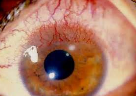
7. Redness: never ignore eye redness. It means there is inflammation and scar tissue will form somewhere if not controlled.

8. Chronic Pain: this is less common but from reasons above.

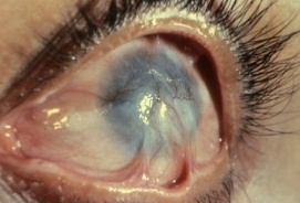
9. Vision Loss: rare. Severe dry eye can lead to vision loss requiring a corneal transplant or even severe eye infections.


Step Ladder Approach to Chronic Eye Itching: 
Allergy Treatments 


Sandra Lora Cremers, MD, FACS: Chronic Eye Itching: Allergy Treatments             
START with STEPS 1, 2 & 3 each day especially if symptoms return. My goal is to arm you with the tools you need when you need them and to teach you when to move to the next tier.

Multi-Tiered, Step Ladder Approach to Allergy TREATMENTS: All the Tools available
  13. Anti-inflammatory pills: low dose steroid or high dose if        severe
12. Autologous serum: available in our office
                   11. Moisture Chamber Glasses: Panoptx, Tranquileyes
 10. Conjunctivochalasis: folding of conjunctiva (clear covering of white part of eye) due to chronic inflammation (other risks: contact lens use, older age, autoimmune thyroid disease) if present, reconstruction of fornix may be needed for relief;
         9. Punctal Plugs (Temporary or Permanent); Probe & Irrigation of
Ducts may be needed; used if itching due to dry eyes
          8. Systemic anti-allergy pills: in conjunction with your PCP: may
need if also have systemic allergic symptoms.
7. If itching is due to Rosacea, we will prescribe: Doxycycline 20mg pill/d:
Avoid Sun; low risk of diarrhea; don’t use if pregnant; If itching is due to
baseline significant dryness, we’ll prescribe: Cyclosporine Restasis
drops: works well; may take weeks to feel benefit
6. Steroid drops: If severe allergy, we recommend a short term low dose
steroid (i.e., Lotemax drops or gel; FML ointment). Use steroids sparingly as chronic
use causes risks: high eye pressure/glaucoma, cataract
5. Allergy Screening test: Takes about 5 minutes to assess 60 common
allergens: If positive, you will know which allergens to avoid & can
consider desensitization &/or discuss with an allergist
4. Rx: If significant itching present, we will prescribe an anti-itch drop —
Lastacaft and Pataday are 1x per day; Bepreve is 2x/day; use for 2 weeks
before allergy season to stabilize cells that cause allergy. OTC Zaditor or Alloway 2x/day helps some. 
3. COLD Artificial tears: put in refrigerator: BEST TO USE Non-preserved but they          can costmore (i.e., Refresh; Oasis tears alternating with Retaine 4-8x/day depending on severity works well). Thicker drops (Soothe, Refresh Dry Eye Therapy) great for severe dryness & itching, though may blur vision; Preserved tears: use no more than 4x per day to avoid allergic reaction to preservative; eyedoc2020.blogspot.comwww.youtube.com à type in search Visionary Eye Doctors for patient education videos 
2. DIET/Life: Goal is to minimize inflammation inside your body: Omega 3’s in Wild Salmon, Fish Oils, Cod Liver Oil: medical grade works best (i.e., PRN 2x/day)
Blink often while working on the computer. Dry eye & allergy symptoms worsen if you blink less. Avoid ceiling fan, direct AC while sleeping at night; or wear a sleep mask. Wear wrap-around sunglasses while in the wind and sun). PersonalHumidifier may help alsoDrink at least 8 glasses (64oz) of filtered or spring water. Avoid intake of reverse osmosis or distilled water due to high acidity & low mineral content; [Recommended, though no randomized studies have proven benefit to date, anti-inflammatory diet (i.e., consider Organic, Paleo Gluten free diet); read books: Grain Brain, Good Calories Bad Calories, Inflammation Nation. Keep doors & windows to outside shut: in spring/summer: use AC when possible (pollen levels increased in open windows/attic fans); Do not hang laundry outside to dry: change clothes, shampoo & shower after being outside when pollen counts are night; Stay indoors during windy weather. Do not mow lawn; Do not rub eyes: if you must rub eyes, wash them first to remove pollen; Keep pets out of bedroom & off bed; Remove all down & feather products from bed, pillows, comforters; Use allergy-free pillow case.

1. Lid Hygiene: helps remove allergens that get caught in the lashes & base of lashes (a daily must in order to improve and prevent symptoms from returning):
(1)   Wash face withhot/warm water (as hot as eyes can stand it without burning skin);
(2)   Apply Warm Compresses 3-5min 2x/d;
(3)    Use Tea Tree Oil (TTO)Cliradex or Avenova daily [may also use TTO shampoo (TTO shampoo in warm water: 1 part in 50) 1x/wk & 50% TTO Daily]: You can cut a piece of Cliradex into pieces with clean scissors and place in Ziploc bag (so it does not dry out); Close eyes & wipe base of eyelashes; allow to air dry 1 minute, wash off before opening eyes; if severe burning, dilute in water. Burning, itching, redness with TTO can last few days: can be allergy or death of demodex mite; decrease or stop if any of above Sx intolerable & let us know.
(4)    Diluted (50%) baby shampoo or Ocusoft Foam: daily if not using TTO or Avenova.
(5)    If no improvement or if immediate improvement needed: will prescribe steroid drops (Lotemax, FML) or ointment (Lotemax, FML, Erythromycin, Bacitracin, Tobradex) at bedtime to apply on lid base. 
Dry Eye Syndrome 
Versus 
Dry Eye Disease: 
What is the Difference 
Between Dry Eye Syndrome 
and Dry Eye Disease?


Dry Eye Syndrome (DES), is the essentially the same as Dry Eye Disease (DED). It is also known as Keratoconjunctivitis Sicca (KCS), and Keratitis Sicca and even sometimes as just, “Dry Eye.” Dry Eye Syndrome is a multifactorial disease of the tears and the ocular surface that results in discomfort which can range from mild irritation to eye burning and go on to terrible pain. It also causes visual disturbances which can begin as blurry vision from time to time and to progress to loss of vision in severe cases. It can cause tearing (usually due to a reflex), redness (which is a sign of inflammation that is leaving behind scar tissue in the cells of the eye), itchiness (which is often due to allergy or demodex mites and/or bacteria on the eyelash margin), and tear film instability (which causes fluctuating vision and the feeling that your glasses are the wrong prescription. All of these issues have the potential to cause significant eye surface damage. 

The most unhappy medical patients I have ever seen are those with cancer with metastatic bone pain. They need iv drips with morphine. Not too far down the list on unhappy patients are my patients with severe dry eye who have terrible pain with each blink of their eye. They ignored their discomfort for years and now regret having ignored their eyes. 


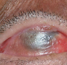
Initially, one or both eyes might be a little red and feel irritated. 

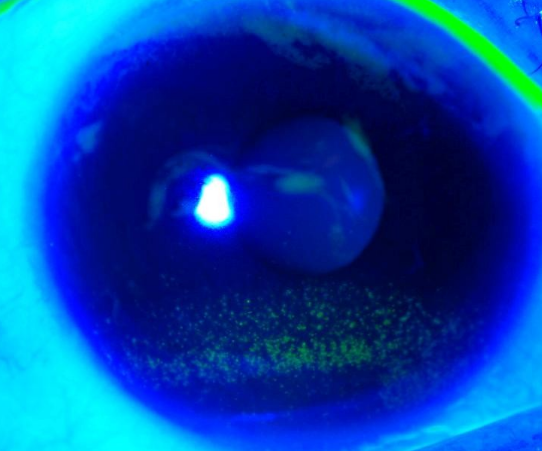
This is what your eyeMD is looking under the microscope.

As the disease worsens, the signs of dry eye under the microscope worsen. This is called Superficial Punctate Keratitis or SPK. This means the cornea’s front layer of cells are dead and not doing their job of protecting the inner nerve layer from discomfort and pain. The front layer or epithelial layer can grow back but they often need help to do so. It is imperative that we help them grow back. Otherwise, more inflammation develops and the disease worsens. It is a vicious cycle that must be stopped.

This is what it looks like when the SPK worsens, demonstrating the worsening of the Dry Eye Disease. 
When the eye senses dryness, it sends a signal which increases inflammation in the form of new blood vessels. These new blood vessels start to grow on the eye. 
In extreme, poorly controlled cases, this is what happens if Dry Eye Syndrome/Dry Eye Disease is left untreated. As you can see, it is potentially sight threatening. And the treatment at this stage is very difficult, very uncomfortable and very expensive.

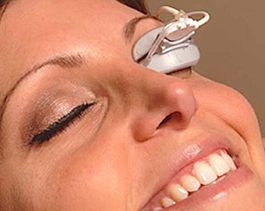
Treatment Options: There are many options. One of the best, we have seen if initial treatments do not resolve symptoms is the Lipiflow machine.

Sandra Lora Cremers, MD, FACS


I revised the Wilkipedia page on its website a bit but was very impressed with the information provided on dry eye.


https://en.wikipedia.org/wiki/Dry_eye_syndrome


Dry eye syndrome

From Wikipedia, the free encyclopedia
Dry eye syndrome
dry eye, keratoconjunctivitis sicca, dry eye disease, keratitis sicca
LG3-LRG.jpg

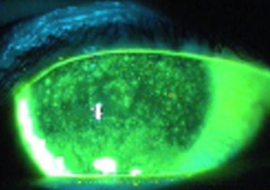
Diffuse lissamine green staining in a person with severe dry eye.[1]
Classification and external resources
Specialty ophthalmology
Dry eye syndrome (DES), also known as keratoconjunctivitis sicca (KCS), is the condition of having dry eyes.[2] Other associated symptoms include irritation, redness, discharge, and easily fatigued eyes. Blurred vision may also occur.[2] The symptoms can range from mild and occasional to severe and continuous.[3] Scarring of the cornea may occur in some cases without treatment.[2]
Dry eye occurs when either the eye does not produce enough tears or when the tears evaporate too quickly.[2] This can result from meibomian gland dysfunctionallergiespregnancySjogren’s syndromevitamin A deficiencyLASIK surgery, and certain medications such as antihistamines, some blood pressure medicationhormone replacement therapy, and antidepressants.[2][3] Chronic conjunctivitis such as from tobacco smoke exposure or infection may also lead to the condition.[2] Diagnosis is mostly based on the symptoms though a number of other tests may be used.[4]
Treatment depends on the underlying cause. Artificial tears are the usual first line treatment. Wrap around glasses that fit close to the face may decrease tear evaporation. Stopping or changing certain medications may help. The medication ciclosporin or steroid eye drops may be used in some cases. Another opinion is lacrimal plugs that prevent tears from draining from the surface of the eye. Dry eyes occasionally makes wearing contact lenses impossible.[2]
Dry eye syndrome is a common eye disease.[3] It affects 5-34% of people to some degree depending on the population looked at.[5] Among older people it affects up to 70%.[6] In China it affects about 17% of people.[7] The phrase “keratoconjunctivitis sicca” means “dryness of the cornea and conjunctiva” in Latin.[8]

Signs and symptoms[edit]

Typical symptoms of dry eye syndrome are dryness, burning[9] and a sandy-gritty eye irritation that gets worse as the day goes on.[10] Symptoms may also be described as itchy, scratchy, stingy or tired eyes.[9][11] Other symptoms are pain, redness, a pulling sensation, and pressure behind the eye.[9][12] There may be a feeling that something, such as a speck of dirt, is in the eye.[9][12] The resultant damage to the eye surface increases discomfort and sensitivity to bright light.[9] Both eyes usually are affected.[13]
There may also be a stringy discharge from the eyes. Although it may seem strange, dry eye can cause the eyes to water. This can happen because the eyes are irritated. One may experience excessive tearing in the same way as one would if something got into the eye. These reflex tears will not necessarily make the eyes feel better. This is because they are the watery type that are produced in response to injury, irritation, or emotion. They do not have the lubricating qualities necessary to prevent dry eye.[12]
Because blinking coats the eye with tears, symptoms are worsened by activities in which the rate of blinking is reduced due to prolonged use of the eyes.[9] These activities include prolonged reading, computer usage, driving, or watching television.[9][12] Symptoms increase in windy, dusty or smoky (including cigarette smoke) areas, in dry environments high altitudes including airplanes, on days with low humidity, and in areas where an air conditioner (especially in a car), fan, heater, or even a hair dryer is being used.[10][9][12][13] Symptoms reduce during cool, rainy, or foggy weather and in humid places, such as in the shower.[9]
Most people who have dry eyes experience mild irritation with no long-term effects. However, if the condition is left untreated or becomes severe, it can produce complications that can cause eye damage, resulting in impaired vision or (rarely) in the loss of vision.[12]
Symptom assessment is a key component of dry eye diagnosis – to the extent that many believe dry eye syndrome to be a symptom-based disease. Several questionnaires have been developed to determine a score that would allow for dry eye diagnosis. The McMonnies & Ho dry eye questionnaire is often used[medical citation needed] in clinical studies of dry eyes.

Causes[edit]

Any abnormality of any one of the three layers of tears produces an unstable tear film, resulting in symptoms of dry eyes.[10]

Decreased tear or excessive evaporation[edit]

Keratoconjunctivitis sicca is usually due to inadequate tear production from lacrimal hyposecretion or to excessive tear evaporation.[10][9] The aqueous tear layer is affected, resulting in aqueous tear deficiency (ATD).[10] The lacrimal gland does not produce sufficient tears to keep the entire conjunctiva and cornea covered by a complete layer.[9] This usually occurs in people who are otherwise healthy. Increased age is associated with decreased tearing.[10] This is the most common type found in postmenopausal women.[9][14]
Causes include idiopathic, congenital alacrimaxerophthalmia, lacrimal gland ablation, and sensory denervation.[10] In rare cases, it may be a symptom of collagen vascular diseases, including relapsing polychondritisrheumatoid arthritisgranulomatosis with polyangiitis, and systemic lupus erythematosus.[10][9][15][16] Sjögren’s syndrome and other autoimmune diseases are associated with aqueous tear deficiency.[10][9] Drugs such as isotretinoin, sedatives, diuretics, tricyclic antidepressantsantihypertensivesoral contraceptives, antihistamines, nasal decongestants, beta-blockers, phenothiazines, atropine, and pain relieving opiates such as morphine can cause or worsen this condition.[10][9][12][13] Infiltration of the lacrimal glands by sarcoidosis or tumors, or postradiation fibrosis of the lacrimal glands can also cause this condition.[10] Recent attention has been paid to the composition of tears in normal or dry eye individuals. Only a small fraction of the estimated 1543 proteins in tears are differentially deficient or upregulated in dry eye, one of which is lacritin.[17][18] Topical lacritin promotes tearing in rabbit preclinical studies.[19] Also, topical treatment of eyes of dry eye mice (Aire knockout mouse model of dry eye) restored tearing, and suppressed both corneal staining and the size of inflammatory foci in lacrimal glands.[20]

Additional causes[edit]

Aging is one of the most common causes of dry eyes because tear production decreases with age.[12] Several classes of medications (both prescription and OTC) have been hypothesized as a major cause of dry eye, especially in the elderly. Particularly, anticholinergic medications that also cause dry mouth are believed to promote dry eye.[21] Dry eye may also be caused by thermal or chemical burns, or (in epidemic cases) by adenoviruses. A number of studies have found that diabetics are at increased risk for the disease.[22][23]
About half of all people who wear contact lenses complain of dry eyes.[12] There are two potential connections between contact usage and dry eye. Traditionally, it was believed that soft contact lenses, which float on the tear film that covers the cornea, absorb the tears in the eyes.[12] However, it is also now known that contact usage damages corneal nerve sensitivity, which subsequently may lead to decreased lacrimal gland tear production and dry eye. The effect of contact on corneal nerve sensitivity is well established for hard contacts as well as soft and rigid gas permeable.[24][25][26] The connection between this loss in nerve sensitivity and tear production is the subject of current research.[27]
Dry eyes also occurs or gets worse after LASIK and other refractive surgeries, in which the corneal nerves are cut during the creation of a corneal flap.[12] The corneal nerves stimulate tear secretion.[12] Dry eyes caused by these procedures usually resolves after several months, but it can be permanent.[13] Persons who are thinking about refractive surgery should consider this.[12]
An eye injury or other problem with the eyes or eyelids, such as bulging eyes or a drooping eyelid can cause keratoconjunctivitis sicca.[11] Disorders of the eyelid can impair the complex blinking motion required to spread tears.[13] Eye injury or disease leading to Boehm Syndrome may be exacerbated by dry eyes.
Abnormalities of the lipid tear layer caused by blepharitis and rosacea, and abnormalities of the mucin tear layer caused by vitamin A deficiency, trachomadiphtheric keratoconjunctivitis, mucocutaneous disorders and certain topical medications are causes of keratoconjunctivitis sicca.[10]
Persons with keratoconjunctivitis sicca have elevated levels of tear nerve growth factor (NGF).[10] It is possible that this ocular surface NGF plays an important role in ocular surface inflammation associated with dry eyes.[10]

Pathophysiology[edit]

Having dry eyes for a while can lead to tiny abrasions on the surface of the eyes.[11] In advanced cases, the epithelium undergoes pathologic changes, namely squamous metaplasia and loss of goblet cells.[10] Some severe cases result in thickening of the corneal surface, corneal erosion, punctate keratopathyepithelial defects, corneal ulceration (sterile and infected), corneal neovascularization, corneal scarring, corneal thinning, and even corneal perforation.[10][9]
Another contributing factor maybe lacritin monomer deficiency. Lacritin monomer, active form of lacritin, is selectively decreased in aqueous deficient dry eye, Sjogren’s syndrome dry eye, contact lens-related dry eye and in blepharitis.[18]

Diagnosis[edit]

Dry eyes can usually be diagnosed by the symptoms alone.[9] Tests can determine both the quantity and the quality of the tears.[13] A slit lamp examination can be performed to diagnose dry eyes and to document any damage to the eye.[10][9]
Schirmer’s test can measure the amount of moisture bathing the eye.[9] This test is useful for determining the severity of the condition.[12] A five-minute Schirmer’s test with and without anesthesia using a Whatman #41 filter paper 5 mm wide by 35 mm long is performed. For this test, wetting under 5 mm with or without anesthesia is considered diagnostic for dry eyes.[10]
If the results for the Schirmer’s test are abnormal, a Schirmer II test can be performed to measure reflex secretion.In this test, the nasal mucosa is irritated with a cotton-tipped applicator, after which tear production is measured with a Whatman #41 filter paper. For this test, wetting under 15 mm after five minutes is considered abnormal.[10]
A tear breakup time (TBUT) test measures the time it takes for tears to break up in the eye.[12] The tear breakup time can be determined after placing a drop of fluorescein in the cul-de-sac.[10]
A tear protein analysis test measures the lysozyme contained within tears. In tears, lysozyme accounts for approximately 20 to 40 percent of total protein content.[10]
A lactoferrin analysis test provides good correlation with other tests.[10]
The presence of the recently described molecule Ap4A, naturally occurring in tears, is abnormally high in different states of ocular dryness. This molecule can be quantified biochemically simply by taking a tear sample with a plain Schirmer test. Utilizing this technique it is possible to determine the concentrations of Ap4A in the tears of patients and in such way diagnose objectively if the samples are indicative of dry eye.[28]
The Tear Osmolarity Test has been proposed as a test for dry eye disease.[29] Tear osmolarity may be a more sensitive method of diagnosing and grading the severity of dry eye compared to corneal and conjunctival staining, tear break-up time, Schirmer test, and meibomian gland grading.[30] Others have recently questioned the utility of tear osmolarity in monitoring dry eye treatment.[18]

Prevention[edit]

There is no way to prevent keratoconjunctivitis sicca. Complications can be prevented by use of wetting and lubricating drops and ointments.[31]

Treatment[edit]

A variety of approaches can be taken to treatment. These can be summarised as: avoidance of exacerbating factors, tear stimulation and supplementation, increasing tear retention, and eyelid cleansing and treatment of eye inflammation.[32]
Dry eyes can be exacerbated by smoky environments, dust and air conditioning and by our natural tendency to reduce our blink rate when concentrating. Purposefully blinking, especially during computer use and resting tired eyes are basic steps that can be taken to minimise discomfort.[32] Rubbing one’s eyes can irritate them further, so should be avoided.[13] Conditions such as blepharitis can often co-exist and paying particular attention to cleaning the eyelids morning and night with mild soaps and warm compresses can improve both conditions.[32]

Environmental control[edit]

Dry, drafty environments and those with smoke and dust should be avoided.[9] This includes avoiding hair dryers, heaters, air conditioners or fans, especially when these devices are directed toward the eyes. Wearing glasses or directing gaze downward, for example, by lowering computer screens can be helpful to protect the eyes when aggravating environmental factors cannot be avoided.[13] Using a humidifier, especially in the winter, can help by adding moisture to the dry indoor air.[9][11][13][32]

Rehydration[edit]

For mild and moderate cases, supplemental lubrication is the most important part of treatment.[10]
Application of artificial tears every few hours[9] can provide temporary relief. Additional research is necessary to determine whether certain artificial tear formulations are superior to others in treating dry eye.[33]

Autologous serum eye drops[edit]

None of the commercially available artificial tear preparations include essential tear components such as epidermal growth factorhepatocyte growth factorfibronectinneurotrophic growth factor, and vitamin A—all of which have been shown to play important roles in the maintenance of a healthy ocular surface epithelial milieu. Autologous serum eye drops contain these essential factors. However, there is some controversy regarding the efficacy of this treatment. At least one study[34] has demonstrated that this modality is more effective than artificial tears in a randomized control study. A 2013 Cochrane review found mixed results when comparing autologous serum eye drops to artificial tears or saline.[35] Evidence from the examined trials showed that autologous serum eye drops resulted in better patient-reported symptoms and improved TBUT, but did not result in improvements in aqueous tear production. No positive effect was observed from ocular surface tests and staining.

Additional options[edit]

Lubricating tear ointments can be used during the day, but they generally are used at bedtime due to poor vision after application.[10] They contain white petrolatummineral oil, and similar lubricants.[10] They serve as a lubricant and an emollient.[10] Application requires pulling down the eyelid and applying a small amount (0.25 in) inside.[10] Depending on the severity of the condition, it may be applied from every hour to just at bedtime.[10] It should never be used with contact lenses.[10] Specially designed glasses that form a moisture chamber around the eye may be used to create additional humidity.[13]

Medication[edit]

Inflammation occurring in response to tears film hypertonicity can be suppressed by mild topical steroids or with topical immunosuppressants such as ciclosporin (Restasis).[36][37]Elevated levels of tear NGF can be decreased with 0.1% prednisolone.[10]
Diquafosol, an agonist of the P2Y2 purinogenic receptor, is approved in Japan for managing dry eye disease by promoting tear secretion.
Lifitegrast is a new drug that was approved by the FDA for the treatment of the condition in 2016.[38]

Fish and Omega−3 fatty acids consumption[edit]

Consumption of dark fleshed fish containing dietary omega-3 fatty acids is associated with a decreased incidence of dry eyes syndrome in women.[39] This finding is consistent with postulated biological mechanisms.[39] Early experimental work on omega-3 has shown promising results when used in a topical application[40] or given orally.[41] A randomized, double-masked study published in 2013 to evaluate the effects of a triglyceride of DHA (Omega-3; Brudy Sec 1.5), showed significant results compared to other methods that are being used.[42]

Cyclosporin[edit]

Topical cyclosporin (topical cyclosporin A, tCSA) 0.05% ophthalmic emulsion is an immunosuppressant.[10] The drug decreases surface inflammation.[13] In a trial involving 1200 people, Restasis increased tear production in 15% of people, compared to 5% with placebo.[12]
It should not be used while wearing contact lenses,[10] during eye infections[12] or in people with a history of herpes virus infections.[13] Side effects include burning sensation (common),[12] redness, discharge, watery eyes, eye pain, foreign body sensation, itching, stinging, and blurred vision.[10][12] Long term use of cyclosporin at high doses is associated with an increased risk of cancer.[43][44]
Cheaper generic alternatives are available in some countries.[45]

Conserving tears[edit]

There are methods that allow both natural and artificial tears to stay longer.[13]
In each eye, there are two puncta[46] — little openings that drain tears into the tear ducts.[12] There are methods to partially or completely close the tear ducts.[13] This blocks the flow of tears into the nose, and thus more tears are available to the eyes.[9] Drainage into either one or both puncta in each eye can be blocked.
Punctal plugs are inserted into the puncta to block tear drainage.[12] For people who have not found dry eye relief with drugs, punctal plugs may help.[12] They are reserved for people with moderate or severe dry eye when other medical treatment has not been adequate.[12]
If punctal plugs are effective, thermal[13] or electric[10] cauterization of puncti can be performed. In thermal cauterization, a local anesthetic is used, and then a hot wire is applied.[13] This shrinks the drainage area tissues and causes scarring, which closes the tear duct.[13]

Other[edit]

Heating systems that try to unblock the oil glands in the eye has some preliminary evidence of benefit.[47]

Surgery[edit]

In severe cases of dry eyes, tarsorrhaphy may be performed where the eyelids are partially sewn together. This reduces the palpebral fissure (eyelid separation), ideally leading to a reduction in tear evaporation.[9]

Prognosis[edit]

Keratoconjunctivitis sicca usually is a chronic problem.[13] Its prognosis shows considerable variance, depending upon the severity of the condition. Most people have mild-to-moderate cases, and can be treated symptomatically with lubricants. This provides an adequate relief of symptoms.[10]
When dry eyes symptoms are severe, they can interfere with quality of life.[12] People sometimes feel their vision blurs with use, or severe irritation to the point that they have trouble keeping their eyes open or they may not be able to work or drive.[9][12]

Epidemiology[edit]

Keratoconjunctivitis sicca is relatively common within the United States, especially so in older patients.[10] Specifically, the persons most likely to be affected by dry eyes are those aged 40 or older.[13] Keratoconjunctivitis sicca is estimated to affect 10% to 20% of adults, with 1 to 4 million aged 65 to 84 affected in the United States.[48]
While persons with autoimmune diseases have a high likelihood of having dry eyes, most persons with dry eyes do not have an autoimmune disease.[13] Instances of Sjögren syndrome and keratoconjunctivitis sicca associated with it are present much more commonly in women, with a ratio of 9:1. In addition, milder forms of keratoconjunctivitis sicca also are more common in women.[10] This is partly because hormonal changes, such as those that occur in pregnancy, menstruation, and menopause, can decrease tear production.[12][13]
In areas of the world where malnutrition is common, vitamin A deficiency is a common cause. This is rare in the United States.[31]
Racial predilections do not exist for this disease.[10]

Synonyms[edit]

Other names for dry eye include dry eye syndrome, keratoconjunctivitis sicca (KCS), dysfunctional tear syndrome, lacrimal keratoconjunctivitis, evaporative tear deficiency, aqueous tear deficiency, and LASIK-induced neurotrophic epitheliopathy (LNE).[2]

Other animals[edit]

Among other animals, keratoconjunctivitis sicca occurs in dogs, cats, and horses.[49]

Dogs[edit]

Keratoconjunctivitis sicca is common in dogs. Most cases are caused by a genetic predisposition, but chronic conjunctivitiscanine distemper, and drugs such as sulfasalazine and trimethoprim-sulfonamide also cause the disease.[50] Symptoms include eye redness, a yellow or greenish discharge, ulceration of the corneapigmented cornea, and blood vessels on the cornea. Diagnosis is made by measuring tear production with a Schirmer tear test. Less than 15 milliliters of tears produced in a minute is abnormal.[50]
Tear replacers are a mainstay of treatment, preferably containing methylcellulose or carboxymethyl cellulose.[50] Ciclosporin stimulates tear production and acts as a suppressant on the immune-mediated processes that cause the disease. Topical antibiotics and corticosteroids are sometimes used to treat secondary infections and inflammation. A surgery known as parotid duct transposition is used in some extreme cases where medical treatment has not helped. This redirects the duct from the parotid salivary gland to the eye. Saliva replaces the tears. Dogs suffering from cherry eye should have the condition corrected to help prevent this disease.
Commonly affected breeds include:

Cats[edit]

Keratoconjunctivitis sicca is uncommon in cats. Most cases seem to be caused by chronic conjunctivitis, especially secondary to feline herpesvirus.[50] Diagnosis, symptoms, and treatment are similar to those for dogs.

See also[edit]

References[edit]

  1. Jump up^ Critser, Brice. “Lissamine green staining in keratoconjunctivitis sicca”Eye Rounds. The University of Iowa. Retrieved 29 July 2016.
  2. Jump up to:a b c d e f g h “Facts About Dry Eye”NEI. February 2013. Retrieved 29 July 2016.
  3. Jump up to:a b c Kanellopoulos, AJ; Asimellis, G (2016). “In pursuit of objective dry eye screening clinical techniques.”Eye and vision (London, England)3: 1. doi:10.1186/s40662-015-0032-4PMC 4716631Freely accessiblePMID 26783543.
  4. Jump up^ Tavares Fde, P; Fernandes, RS; Bernardes, TF; Bonfioli, AA; Soares, EJ (May 2010). “Dry eye disease.”. Seminars in ophthalmology25 (3): 84–93. doi:10.3109/08820538.2010.488568PMID 20590418.
  5. Jump up^ Messmer, EM (30 January 2015). “The pathophysiology, diagnosis, and treatment of dry eye disease.”Deutsches Arzteblatt international112 (5): 71–81; quiz 82. doi:10.3238/arztebl.2015.0071PMC 4335585Freely accessiblePMID 25686388.
  6. Jump up^ Ding, J; Sullivan, DA (July 2012). “Aging and dry eye disease.”Experimental Gerontology47 (7): 483–90. doi:10.1016/j.exger.2012.03.020PMC 3368077Freely accessiblePMID 22569356.
  7. Jump up^ Liu, NN; Liu, L; Li, J; Sun, YZ (2014). “Prevalence of and risk factors for dry eye symptom in mainland china: a systematic review and meta-analysis.”Journal of ophthalmology2014: 748654. doi:10.1155/2014/748654PMC 4216702Freely accessiblePMID 25386359.
  8. Jump up^ Firestein, Gary S. (2013). Kelley’s textbook of rheumatology (9th ed.). Philadelphia, PA: Elsevier/Saunders. p. 1179. ISBN 9781437717389.
  9. Jump up to:a b c d e f g h i j k l m n o p q r s t u v w x y “Keratoconjunctivitis Sicca”The Merck Manual, Home EditionMerck & Co., Inc. 2003-02-01. Retrieved 2006-11-12.
  10. Jump up to:a b c d e f g h i j k l m n o p q r s t u v w x y z aa ab ac ad ae af ag ah ai aj ak al“Keratoconjunctivitis, Sicca”eMedicineWebMD, Inc. January 27, 2010. Retrieved September 3, 2010.
  11. Jump up to:a b c d “Dry eyes”MedlinePlus Medical EncyclopediaU.S. National Library of Medicine. 2006-10-04. Retrieved 2006-11-16.
  12. Jump up to:a b c d e f g h i j k l m n o p q r s t u v w x y z Michelle Meadows (May–June 2005). “Dealing with Dry Eye”FDA Consumer MagazineU.S. Food and Drug Administration. Archived from the original on February 23, 2008.
  13. Jump up to:a b c d e f g h i j k l m n o p q r s t u “Dry eyes”Mayo Clinic. Mayo Foundation for Medical Education and Research. 2006-06-14. Retrieved 2006-11-17.
  14. Jump up^ Sendecka M, Baryluk A, Polz-Dacewicz M (2004). “Częstość występowania zespołu suchego oka” [Prevalence and risk factors of dry eye syndrome]Przegla̧d Epidemiologiczny (in Polish). 58 (1): 227–33. PMID 15218664.
  15. Jump up^ Puéchal, X; Terrier, B; Mouthon, L; Costedoat-Chalumeau, N; Guillevin, L; Le Jeunne, C (March 2014). “Relapsing polychondritis.”. Joint, bone, spine : revue du rhumatisme81 (2): 118–24. doi:10.1016/j.jbspin.2014.01.001PMID 24556284.
  16. Jump up^ Cantarini, L; Vitale, A; Brizi, MG; Caso, F; Frediani, B; Punzi, L; Galeazzi, M; Rigante, D (February–March 2014). “Diagnosis and classification of relapsing polychondritis.”. Journal of autoimmunity. 48-49: 53–9. doi:10.1016/j.jaut.2014.01.026PMID 24461536.
  17. Jump up^ Zhou L, Zhao SZ, Koh SK, et al. (July 2012). “In-depth analysis of the human tear proteome”. Journal of Proteomics75 (13): 3877–85. doi:10.1016/j.jprot.2012.04.053PMID 22634083.
  18. Jump up to:a b c Karnati R, Laurie DE, Laurie GW (December 2013). “Lacritin and the tear proteome as natural replacement therapy for dry eye”Experimental Eye Research117: 39–52. doi:10.1016/j.exer.2013.05.020PMC 3844047Freely accessiblePMID 23769845.
  19. Jump up^ Samudre S, Lattanzio FA, Lossen V, et al. (August 2011). “Lacritin, a novel human tear glycoprotein, promotes sustained basal tearing and is well tolerated”Investigative Ophthalmology & Visual Science52 (9): 6265–70. doi:10.1167/iovs.10-6220PMC 3176019Freely accessiblePMID 21087963.
  20. Jump up^ Vijmasi T, Chen FY, Balasubbu S, Gallup M, McKown RL, Laurie GW, McNamara NA (July 2014). “Topical administration of lacritin is a novel therapy for aqueous-deficient dry eye disease.”Invest Ophthalmol Vis Sci55 (8): 5401–9. doi:10.1167/iovs.14-13924PMC 4148924Freely accessiblePMID 25034600.
  21. Jump up^ Fraunfelder FT, Sciubba JJ, Mathers WD (2012). “The role of medications in causing dry eye”Journal of Ophthalmology2012: 285851. doi:10.1155/2012/285851PMC 3459228Freely accessiblePMID 23050121.
  22. Jump up^ Kaiserman I, Kaiserman N, Nakar S, Vinker S (March 2005). “Dry eye in diabetic patients”. American Journal of Ophthalmology139 (3): 498–503. doi:10.1016/j.ajo.2004.10.022PMID 15767060.
  23. Jump up^ Li H, Pang G, Xu Z (2004). “Tear film function of patients with type 2 diabetes”. Zhongguo Yi Xue Ke Xue Yuan Xue Bao26 (6): 682–6. PMID 15663232.
  24. Jump up^ Millodot M (1978). “Effect of long term wear of hard contact lenses on corneal sensitivity”. Arch Ophthalmol96 (7): 1225–7. doi:10.1001/archopht.1978.03910060059011PMID 666631.
  25. Jump up^ Macsai MS, Varley GA, Krachmer JH (1990). “Development of keratoconus after contact lens wear. Patient characteristics”. Arch Ophthalmol108 (4): 534–8. doi:10.1001/archopht.1990.01070060082054PMID 2322155.
  26. Jump up^ Murphy PJ, Patel S, Marshall J (2001). “The effect of long term daily contact lens wear on corneal sensitivity”. Cornea20 (3): 264–9. doi:10.1097/00003226-200104000-00006PMID 11322414.
  27. Jump up^ Mathers WD, Scerra C (2000). “Dry eye; investigators look at syndrome with new model”. Ophthalmol Times25 (7): 1–3.
  28. Jump up^ Peral A, Carracedo G, Acosta MC, Gallar J, Pintor J (September 2006). “Increased levels of diadenosine polyphosphates in dry eye”. Investigative Ophthalmology & Visual Science47 (9): 4053–8. doi:10.1167/iovs.05-0980PMID 16936123.
  29. Jump up^ Tomlinson, A (April 2007). “DEWS Report, Ocular Surface April 2007 Vol 5 No 2” (PDF).
  30. Jump up^ “American Academy of Ophthalmology Cornea/External Disease Panel. 2011 Preferred Practice Pattern® (PPP) guidelines”. October 2011.
  31. Jump up to:a b “Dry eyes syndrome”MedlinePlus Medical EncyclopediaU.S. National Library of Medicine. 2006-10-04. Retrieved 2006-11-16.
  32. Jump up to:a b c d Lemp MA. (2008). “Management of Dry Eye”American Journal of Managed Care14 (4): S88–S101. PMID 18452372. Retrieved 2008-07-25.
  33. Jump up^ Pucker AD, Ng SM, Nichols JJ (2016). “Over the counter (OTC) artificial tear drops for dry eye syndrome”. Cochrane Database Syst Rev2: CD009729. doi:10.1002/14651858.CD009729.pub2PMID 26905373.
  34. Jump up^ Kojima, T.; Higuchi, A.; Goto, E.; Matsumoto, Y.; Dogru, M.; Tsubota, K. (2008). “Autologous Serum Eye Drops for the Treatment of Dry Eye Diseases”. Cornea27: S25–S30. doi:10.1097/ICO.0b013e31817f3a0ePMID 18813071.
  35. Jump up^ Pan Q, Angelina A, Zambrano A, Marrone M, Stark WJ, Heflin T, Tang L, Akpek EK (2013). “Autologous serum eye drops for dry eye”Cochrane Database Syst Rev10: CD009327. doi:10.1002/14651858.CD009327.pub2PMC 4007318Freely accessiblePMID 23982997.
  36. Jump up^ Tatlipinar S, Akpek E (2005). “Topical cyclosporine in the treatment of ocular surface disorders”Br J Ophthalmol89 (10): 1363–7. doi:10.1136/bjo.2005.070888PMC 1772855Freely accessiblePMID 16170133.
  37. Jump up^ Barber L, Pflugfelder S, Tauber J, Foulks G (2005). “Phase III safety evaluation of cyclosporine 0.1% ophthalmic emulsion administered twice daily to dry eye disease patients for up to 3 years”. Ophthalmology112 (10): 1790–4. doi:10.1016/j.ophtha.2005.05.013PMID 16102833.
  38. Jump up^ “FDA approves new medication for dry eye disease”FDA. July 12, 2016. Retrieved 13 July2016.
  39. Jump up to:a b Miljanović B, Trivedi KA, Dana MR, Gilbard JP, Buring JE, Schaumberg DA (2005). “Relation between dietary n-3 and n-6 fatty acids and clinically diagnosed dry eye syndrome in women”Am J Clin Nutr82 (4): 887–93. PMC 1360504Freely accessiblePMID 16210721.
  40. Jump up^ Rashid S, Jin Y, Ecoiffier T, Barabino S, Schaumberg M, Dana RD (2008). “Topical Omega-3 and Omega-6 Fatty Acids for Treatment of Dry Eye”. Arch Ophthalmol126 (2): 219–225. doi:10.1001/archophthalmol.2007.61PMID 18268213.
  41. Jump up^ Creuzot C, Passemard M, Viau S, Joffre C, Pouliquen P, Elena PP, Bron A, Brignole F (2008). “Improvement of dry eye symptoms with polyunsaturated fatty acids”. J Fr Ophthalmol29 (8): 868–73. PMID 17075501.
  42. Jump up^ Oleñik A, Jiménez-Alfaro I, Alejandre-Alba N, Mahillo-Fernández I (2013). “A randomized, double-masked study to evaluate the effect of omega-3 fatty acids supplementation in meibomian gland dysfunction”Clinical Interventions in Aging8: 1133–8. doi:10.2147/CIA.S48955PMC 3770496Freely accessiblePMID 24039409.
  43. Jump up^ “Restasis” (PDF)Allergan. January 2008. Retrieved 2008-07-23.
  44. Jump up^ Dantal J, Hourmant M, Cantarovich D, Giral M, Blancho G, Dreno B, Soulillou JP (1998). “Effect of long-term immunosuppression in kidney-graft recipients on cancer incidence: randomised comparison of two cyclosporin regimens”. The Lancet351 (9103): 623–628. doi:10.1016/S0140-6736(97)08496-1PMID 950031760 patients developed cancers, 37 in the normal-dose group and 23 in the low-dose group (p<0.034); 66% were skin cancers (26 vs 17; p<0.05). The low-dose regimen was associated with fewer malignant disorders but more frequent rejection.
  45. Jump up^ “Sun Pharma Product List”. Sun Pharma. Archived from the original on 2007-02-13. Retrieved 2006-11-27.
  46. Jump up^ Carter SR (June 1998). “Eyelid disorders: diagnosis and management”American Family Physician57 (11): 2695–702. PMID 9636333.
  47. Jump up^ Qiao, J; Yan, X (2013). “Emerging treatment options for meibomian gland dysfunction.”Clinical ophthalmology (Auckland, N.Z.)7: 1797–803. doi:10.2147/OPTH.S33182PMC 3772773Freely accessiblePMID 24043929.
  48. Jump up^ Ervin AM, Wojciechowski R, Schein O (2010). “Punctal occlusion for dry eye syndrome”Cochrane Database Syst Rev9: CD006775. doi:10.1002/14651858.CD006775.pub2PMC 3729223Freely accessiblePMID 20824852.
  49. Jump up^ “Keratoconjunctivitis, Sicca”The Merck Veterinary ManualMerck & Co., Inc. Retrieved 2006-11-18.
  50. Jump up to:a b c d e Gelatt, Kirk N. (ed.) (1999). Veterinary Ophthalmology (3rd ed.). Lippincott, Williams & Wilkins. ISBN 0-683-30076-8.

Further reading[edit]

External links[edit]

Shopping Cart