Keeping pace with this vigorous progress, physicians, surgeons, academic centers, hospitals, and private offices across the world are continually updating their equipment and services in order to offer the latest in high-tech surgical and medical care. From specialty surgical and medical centers with state-of-the-art digital imaging scanners, to multi-disciplinary mega-hospitals featuring cutting-edge medical robots, all surgeons and physicians are trying to provide the best possible care with the best efficiency… since every one of us, especially a sick patient, hates waiting and waiting to see the doctor or doctor’s staff.
Everyone I know is working hard on this issue. Still we have emergencies that interrupt a perfectly orchestrated schedule to minimize our waiting times.
In ophthalmology we are blessed and cursed. The eye is so intricate that we now have practically a machine for each of it’s important layers and functions. At Harvard, each of the key machines was on a different floor. The Retina floor had the OCT. The Glaucoma department had the HRT, HVF. The Cornea department had the only MARCO in the whole institution and there were many fights as to who could use it.
In private practice, we are blessed to have all the key machines in one location. Still because imaging the many areas of the eye are crucial in many patients to being sure one does not have a potentially blinding or debilitating chronic pain condition, it can take hours to get through an eye doctor’s basic visit.
This is frustrating for everyone.
Also, no one wants to be made to feel they are on a conveyor belt of testing. But this is what it seems we have to deal with.
Currently patients check in and then sit down. They they are called to have basic testing. They have to get up and sit down at each of the following machines at times. If the doctor needs another test, they have to go back and get the other machine check. Each machine is crucial for particular issues and sometimes the patient will not complain about an issue unless asked point blank by the MD and then the machine can help determine if further testing or surgery is needed.
Our biggest obstacle is how to evaluate all the layers of the eye as efficiently as possible without making the patient feel they are literally on a conveyor belt.
I love these machines! They make me the best physician and surgeon I can be. I have saved countless lives and patients’ eyesights with these machines! When I walked into Visionary Eye Doctors for the first time after having been at Harvard for 9 years, my jaw dropped! “You have all these machines in one floor? Are you kidding me!” I was so thrilled! I still am.
A few months ago, a 48 year old patient came in to the front desk and announced, “I think I am having a stroke.” But he could move his hands and arms and walk and talk normally. His vision was normal 20/20. He was so sure of it, we did an immediate Visual Field (below machine) and sure enough he was right! He was having the beginnings of a stroke. The ambulance came and he had immediate care at the hospital nearby and came in with tears to thank me recently for saving his life. If he had had to wait to get the visual field, the story might have ended differently.
Below:
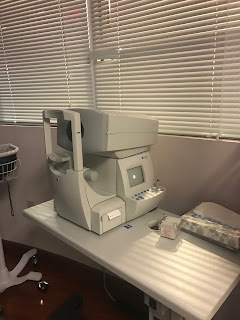
This is the autorefractor that allows us to more precisely determine what glasses Rx a patient might need. It is sometimes repeated after dilation to determine if headaches or eye strain are due to latent hyperopia (needing a stronger glasses Rx.


This is the MARCO Refraction System. This is one of the most expensive machines out there. It can do an autorefraction and check your need for glasses Rx fast. The machine in the back measures your current glasses Rx.


This is a Humphrey Visual Field (HVF): this determines if there is a defect in the optical pathway of the brain, from the occipital cortex to the optic nerve. It helps us determine if one had glaucoma (localized nerve damage) or a brain tumor.

These are the ERG and VEP machines that allow us to determine the functional health of the retina and optic nerve. If the MD cannot see anything abnormal on the microscope, these machines can pick up sub-clinical diseases. These are used for retina and optic nerve diseases.


