Neruopathic Eye Pain: What is it and how should it be treated?


Patients that have “Pain without Stain” or “Neuropathic Dry Eye” are often our toughest patients to help. Patients remain frustrated as most standard dry eye remedies do not relieve all the pain or do nothing to help. Doctors/surgeons/doctors remain frustrated as all their recommendations and treatments do not fully heal the patient. Many eye surgeons have given up on even seeing these patients for this reason. 


A colleague noted recently that the temptation for eye surgeons is to limit one’s practice to only surgical patients and “not waste chair-time” by these “impossible to treat patients.” Unfortunately many eye surgeons have this sentiment. 


Many of these patients have tried all standard options provide. These patients come to see me usually having tried multiple options with no relief and having seen many eye doctors and surgeons on the way to see me. Some of these patients are helped significantly with Intense Pulse Light and Meibomian Gland Probing/Expression with or without Autologous Serum, PRP, and (we await results for Stem Cell injections). Some of them have not been helped with even these procedures. 


What is the cause of Neuropathic Eye Pain/Neuropathic Dry Eye?


Neuropathic pain/ Neuropathic Dry eye is due to a combination of many factors. Chronic inflammation is a key component. There are many factors that can contribute to chronic, microscopic inflammation at the nerve cell level.


Prolonged ocular surface stimulation can alter neurons and lead to pain amplification expressed chonic eye pain. My concern is that the chronic stimulation of looking at electronic screens is a big factor that is taking many patients over the edge. Other negative factors that cause chronic microscopic inflammation are previous isotretinoin/Accutane use, LASIK/PRK, chronic allergy, ocular rosacea, oral contraceptives use [Recently a patient told me a world renowned Harvard eye surgeon told her that her Mirena IUD caused her Sjogren’s syndrome: more on that in an upcoming post], smoking, previous eye trauma, autoimmune disease (like Sjogren’s syndrome, Rheumatoid Arthritis, Lupus), previous chemotherapy, radiation, Bells Palsy, Stevens Johnson Syndrome, Graft Versus host disease. Also general aging and hormonal changes can cause nerve fiber irregularities which can also exacerbate chronic eye pain symptoms. 


Many patients with Neuropathic pain/ Neuropathic Dry eye also have other disorders that central pain processing, such as depression, anxiety, and insomnia. (Reference 2, 3)


Inflammation begins on the ocular surface with chronic stimulants leading to chronic inflammatory cytokines release. This can lead to increased activity of sensory nerves or chronic sensory nerve firing. Over time, chronic microscopic inflammation can lead to peripheral sensitization which means the corneal nerves become “numb” to the stimulus and continue to fire even without stimuli or irritants. 


As Dr. Anat Galor notes in her paper below (Reference 1):
Increased activity of sensory nerves can also cause inflammation in the form of neuro-inflammation, which in turn ramps up the activity of pain-carrying nerves, leading to a self-perpetuating phenomenon known as peripheral sensitization.7 For many patients this can be reversed after resolution of the inciting stress and ocular surface inflammation.8 Some dry-eye patients experience only transient episodes of pain, whereas others have persistent symptoms of chronic disease. 


Neuropathic pain is by definition “pain arising as direct consequence of a lesion or disease affecting the somatosensory system,” and is often chronic.9


The transmission of dry-eye pain signals to the somatosensory cortex is not a passive process. Along the way, these electrical signals are modified by feed-forward and feedback systems that typically intensify the signals. This explains the unique property of pain to become amplified during a constant noxious stimulus, in contrast to most other types of sensory responses that adapt during persistent stimulation and thereby attenuate the responses. This physiological phenomenon, known as central sensitization, occurs with dry eye-like pain as well.8 Since the trigeminal brainstem was shown in animal models to be the location of central control of homeostatic corneal wetness,10 corneal algesia11 and aversive responses to light,12 the brainstem may also be the origin of the clinical expression of dry-eye-related pain. The possibility that disorders of the dry-eye alarm system itself can explain the variety of clinical patterns associated with dry eye-like pain offers a strikingly different perspective. Moreover, the location and persistence of this dysfunctional alarm system can alter the functional anatomy through the well-known, innately powerful neuroplasticity of the central nervous system. These maladaptive changes in the CNS result in neuropathic pain.

Possible Origins of the Pain

For us to be able to manage these cases effectively, it’s important to recognize the factors influencing neuropathic pain. Age-related dry-eye disease is characterized by the attrition of corneal nerve fibers. Its consequences are associated with an increased sensitivity to tear evaporation, or corneal evaporative hyperalgesia. Pain fiber attrition also occurs in de-afferentation hypersensitivity, a phenomenon found in the skin of healthy elderly subjects. The loss of nerve fibers in these conditions is associated with increased activity of the surviving nerves.13 The parallels between this condition and age-related dry eye are striking. 

In mice, it has been demonstrated that sensory nerve injuries, such as those caused by LASIK axotomies, trigger a phenotypic change in the somata of the surviving nerves from conduction to regeneration, promoting the expression of atopic pain generators that are hypersensitive and hyper-responsive14 and that are transported to the regenerating nerve sprouts and the central terminals of severed axons from the nerve somata in the trigeminal ganglion where they’re expressed. This likely is responsible for the complaints of dry-eye symptoms following refractive procedures. Persistence of the regenerating phenotype long after healing has occurred, sometimes years after LASIK, as suggested by the characteristic morphology of regenerating nerves and increased numbers of mature dendritic cells in the sub-basal plexus, may explain the chronicity of dry-eye pain even in the absence of external signs of inflammation.


Both nociceptive and neuropathic mechanisms likely play a role in chronic itch and DE. Nociceptive pain (defined as pain that arises from actual or threatened damage to non-neural tissue and is due to the activation of nociceptors) is driven by chronic ocular surface abnormalities that are seen in many DE sub-types (pSS, graft versus host disease). With time, inflammation, trauma, and chronic activation of nociceptors can lead to permanent changes in nociceptor structure and function (i.e. sensitization) that can occur both peripherally and/or centrally, thus adding a neuropathic component to the pain (defined as a pain due to a lesion or disease of the somatosensory system).”


Thus there are patients who have tried many of the standard options but still have chronic pain. 


It is very frustrating to not be able to provide comfort to a suffering patient.


More and more of my severe dry eye patients are trying these oral drugs below. 

Most, unfortunately have not helped or the patient could not tolerate the side effects, but some cannot live without them. 


Why do some patients get better with these pills and others do not? No one knows for sure, but in some patients it is worth a try if all else has been tried. 


How do you tell if you have neuropathic pain?

Here are some ways we can tell the eye pain is Neuropathic:
1. The eye pain remains the same after the installation of anesthetic. Those patients without neuropathic pain get relief of their pain usually. 
2. There are no real objective signs of dry eye on the cornea or minimal meibomian glands atrophy.
3. Take the Neuropathic Pain Symptom Inventory. 
Dr. Anat Galor adds in these questions: See below
a. Do your eyes feel hot and burning?
b. Are you sensitivity to light?
c. Are you sensitivity to wind?


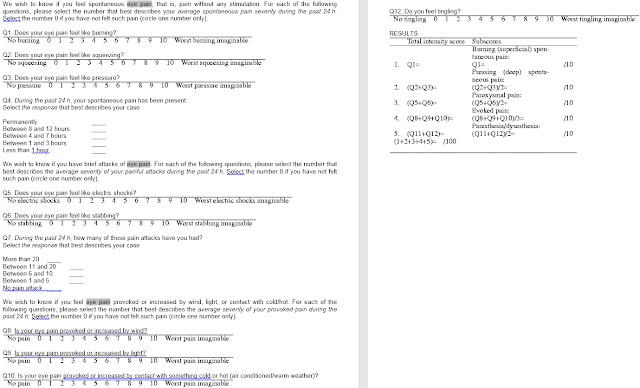
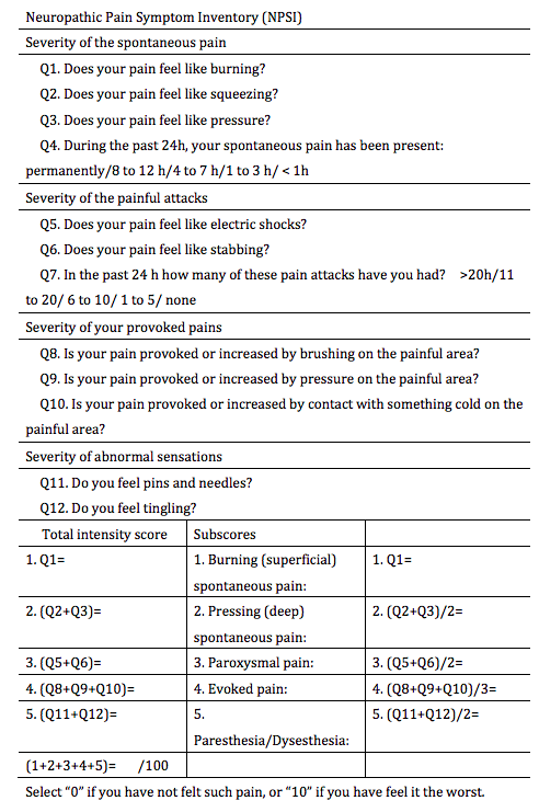
Dr. Anat Galor’s Version of Neuropathic Pain Symptom Inventory (NPSI): from reference 1 below & posted here with her permission. 


“The NPSI has been validated as a self-report instrument for assessing neuropathic pain, has been used to quantify different aspects of neuropathic pain, and has been found to correlate with mechanical and/or thermal allodynia and hyperalgesia assessed using Quantitative Sensory Testing (QST).


Original


How to Treat Neuropathic Eye Pain?

1. First try all the standard options for dry eye pain: Lid hygiene, warm/hot compresses with lid massaging, blinking exercises, frequent blinking, long-paused blinking, voiding electronic screen use as much as possible (also take breaks every 10min for 10 sec, look 10f away), clean eyelids with diluted tea tree oil and/or Avenova or hydrochor; use only non-preserved artificial tears (Retaine or Omega rich tears), Eat Omega 3 2000-4000mg/day; stay on an anti-inflammatory diet until we can get the pain to go away (ie, gluten free, Paleo recommended); Use Doxycyline 20mg/day if able to tolerate and/or have ocular rosacea as a contributing cause. 
Get meibography and have it scored.
2. If these do not help and you have enough oil glands on meibography, try Lipiflow at least 2x over a 6-10 month period (this is the only Dry Eye procedure approved by the FDA to save meibomian glands but only 70% or so of patients get relief of dry eye symptoms). 
3. If no improvement: try IPL if you are a candidate with videotaped expression x 4-6 (if skin type allows this) to see if this helps.
4. If still no improvement, try Autologous Serum (AS), or Platelet rich plasma (PRP) eye drops. Amniotic drops have come out but no publications yet say it helps unlike AS, PRP to my knowledge. 
5. Meibomian Gland Probing and Expression first alone and then with Autologous Serum (AS), or Platelet rich plasma (PRP) if AS did not help has helped many patients with Neuropathic Eye Pain but it is not 100%. Studies into the use of autologous Stem Cell injections into the meibomian glands, lacrimal glands, subconjunctival, and intravenous have been started but no publications have shown they work yet. All these treatments in #5 are experimental and not FDA approved.
6. Try also Prokera/Amniotic membrane to see if this helps though this can be an uncomfortable procedure that lasts 4-5days. 

7. Oral medications:  If all of this fails, I would consider trying one of the below drugs. These have higher risks and side effects than above options in general. They are not FDA approved to use in Dry Eye so are being prescribed “off label.” 


Some patients may respond to drugs, such as:

a. Naltrexone,
b. Neurontin (gabapentin, Pfizer), 
c. Lyrica (pregabalin, Pfizer) 
d. Cymbalta (duloxetine, Eli Lilly)

e. Nortriptyline


The key issue is that there are no published studies tha I could find to date demonstrating a relief of neuropathic dry eye pain compared to placebo or controls on any of these medications. 


The most common one tried first is Naltrexone. Some surgeons prescribe Natrexone along with nortriptyline but there have been no published papers to prove any of these drugs (nor their combination) help above placebo. 


Many of these have significant side effects, such as making one suicidal. I had another young patient come in on Friday saying Naltrexone made her want to jump off a bridge. Why does this happen to some and not to others? 


The key side effects I have heard from patients on these drugs is the disturbance of sleep, nightmares, and suicidal ideation. It is important if you are on these medications for dry eye, you tell your family and keep an open dialog with your loved ones and your prescriber if you have any of these side effects. 


Safer treatments to try for Neuropathic Dry Eye, which have shown positive improvements are:

1. Autologous Serum
2. Platelet rich plasma.


But most of the patients I have seen on the oral drugs have tried these and they have not work.


We await the results to see if Autologous Stem Cell injections (with or without a combination of PRP) works better than any of the options above.


For now, it is helpful to know you have a component of neuropathic pain and know you are not crazy. It is important to be treated for any associated or underlying anxiety or depression from the pain and have a pain specialist follow you.


A recent patient told me the only place he finds relief from his pain is in a very hot (dry not wet) sauna. Nothing else helps. He has tried some of the oral meds above and they did not help.  Only the hot sauna helped.


Why? I do not know, but there must be a way to replicate that pain relief without having to live in a hot sauna. 


SLC


References:


1. 

 2018 Jan 17;115:T5. eCollection 2017 Aug.

The Relationship Between Ocular Itch, Ocular Pain, and Dry Eye Symptoms (An American Ophthalmological Society Thesis).

Abstract

PURPOSE:

To evaluate associations between sensations of ocular itch and dry eye (DE) symptoms, including ocular pain, and DE signs.

METHODS:

A cross-sectional study of 324 patients seen in the Miami Veterans Affairs eye clinic was performed. The evaluation consisted of questionnaires regarding ocular itch, DE symptoms, descriptors of neuropathic-like ocular pain (NOP), and evoked pain sensitivity testing on the forehead and forearm, followed by a comprehensive ocular surface examination including corneal mechanical sensitivity testing. Analyses were performed to examine for differences between those with and without subjective complaints of ocular itch.

RESULTS:

The mean age was 62 years with 92% being male. Symptoms of DE and NOP were more frequent in patients with moderate-severe ocular itch compared to those with no or mild ocular itch symptoms. With the exception of ocular surface inflammation (abnormal matrix metalloproteinase 9 testing) which was less common in those with moderate-severe ocular itch symptoms, DE signs were not related to ocular itch. Individuals with moderate-severe ocular itch also demonstrated greater sensitivity to evoked pain on the forearm and had higher non-ocular pain, depression, and post-traumatic stress disorders scores, compared to those with no or mild itch symptoms.

CONCLUSIONS:

Subjects with moderate-severe ocular itch symptoms have more severe symptoms of DE, NOP, non-ocular pain and demonstrate abnormal somatosensory testing in the form of increased sensitivity to evoked pain at a site remote from the eye, consistent with generalized hypersensitivity.

PMID:


29391860


PMCID:


PMC5774848


[Indexed for MEDLINE] 


Free PMC Article


REFERENCE:
2. Galor A, Feuer W, Lee DJ, et al. Depression, post-traumatic stress disorder, and dry eye syndrome: a study utilizing the national United States Veterans Affairs administrative database. Am J Ophthalmol. 2012 Aug;154(2):340–346 e342.
3. Labbe A, Wang YX, Jie Y, Baudouin C, Jonas JB, Xu L. Dry eye disease, dry eye symptoms and depression: the Beijing Eye Study. Br J Ophthalmol. 2013 Nov;97(11):1399–1403.


Some information on Lyrica


• Begin dosing at 150 mg/day
• Administer in 2 or 3 divided doses per day
• May be increased to 300 mg/day within 1 week
• Maximum dose of 600 mg/day.
Dose should be adjusted in patients with reduced renal function. (2.5)

Most common adverse reactions (≥ 5% and twice placebo) are dizziness, somnolence, dry mouth, edema, blurred vision, weight gain and thinking abnormal (primarily difficulty with concentration/attention). 

• Angioedema (e.g. swelling of the throat, head and neck) can occur, and may be associated with life-threatening respiratory compromise requiring emergency treatment. Discontinue LYRICA immediately in these cases. 

• Hypersensitivity reactions (e.g. hives, dyspnea, and wheezing) can occur. Discontinue LYRICA immediately in these patients. (5.2)

If experienced, these tend to have a Severe expression
  • Confused
  • Decreased Blood Platelets
  • Infection
  • Inflammation Of The Lining Of The Stomach And Intestines
  • Puffy Face From Water Retention
  • Sensation Of Spinning Or Whirling
If experienced, these tend to have a Less Severe expression
  • Constipation
  • Extreme Sense Of Well Being
  • Feeling Weak
  • Head Pain
  • Injury
  • Sinus Irritation And Congestion
  • Stomach Cramps
  • Twitching

RARE side effects


If experienced, these tend to have a Severe expression
  • Chest Pain
  • Difficulty Speaking
  • Double Vision
  • Dyskinesia
  • Giant Hives
  • Having Thoughts Of Suicide
  • Hemorrhage Of Blood Under The Skin
  • Hives
  • Low Blood Sugar
  • Middle Ear Infection
  • Muscle Spasm
  • Pink Eye
  • Problem Behavior
  • Problems With Bladder Control
  • Prolonged P-R Interval Observed On EKG
  • Rash
  • Ringing In The Ears
  • Small Skin Blister
  • Trouble Breathing
  • Visible Water Retention
  • Wheezing
If experienced, these tend to have a Less Severe expression
  • Abnormal Increase In Muscle Tone
  • Abnormal Manner Of Walking
  • Altered Interest In Having Sexual Intercourse
  • Backache
  • Bronchitis
  • Diarrhea
  • Enlarged Breasts
  • Feel Like Throwing Up
  • Feeling Anxious
  • Fever
  • Fingernail And/Or Toenail Disease
  • Flu-Like Symptoms
  • Frequent Urination
  • Gas
  • Inability To Have An Erection
  • Increased Hunger
  • Involuntary Eye Movement
  • Involuntary Quivering
  • Itching
  • Joint Pain
  • Leg Cramps
  • Loss Of Memory
  • Loss Of One’s Own Sense Of Reality Or Identity
  • Low Energy
  • Muscle Pain
  • Muscle Weakness
  • Nervous
  • Numbness
  • Problems With Eyesight
  • Stupor
  • Swelling Of The Abdomen
  • Throwing Up

—-
7.
There is no proof published that I can find to date proving gabapentin as a favorable treatment in neuropathic dry eye pain. Still I have many patients that say it works.
Below is the only paper published on Pubmed when I query, “gabapentin neuropathic eye pain.”


1.
Wang TX, Yin D, Guo W, Liu YY, Li YD, Qu WM, Han WJ, Hong ZY, Huang ZL.
Pharmacol Biochem Behav. 2015 Aug;135:31-9. doi: 10.1016/j.pbb.2015.05.007. Epub 2015 May 16.

PMID:


25989046

2.
Stidd DA, Wuollet AL, Bowden K, Price T, Patwardhan A, Barker S, Weinand ME, Annabi J, Annabi E.
Pain Physician. 2012 Jan-Feb;15(1):27-33.

PMID:


22270735


Free PMC Article

3.
Takemura Y, Yamashita A, Horiuchi H, Furuya M, Yanase M, Niikura K, Imai S, Hatakeyama N, Kinoshita H, Tsukiyama Y, Senba E, Matoba M, Kuzumaki N, Yamazaki M, Suzuki T, Narita M.
Synapse. 2011 Jul;65(7):668-76. doi: 10.1002/syn.20898. Epub 2011 Feb 25.

PMID:


21162109

4.
Yennurajalingam S, Peuckmann V, Bruera E.
Support Cancer Ther. 2004 Jan 1;1(2):97-110. doi: 10.3816/SCT.2004.n.003.

PMID:


18628186

5.
Kavalieratos CS, Dimou T.
Pain Med. 2008 Apr;9(3):377-8. doi: 10.1111/j.1526-4637.2006.00167.x.

PMID:


18366517

6.
Sloan PA, Kancharla A.
Pain Palliat Care Pharmacother. 2003;17(2):89-94.

PMID:


14649392

———–


8.
When I query: “gabapentin dry eye,” only this comes up. 

 2010 Jul;81(7):731-3. doi: 10.1136/jnnp.2009.177303.


Non-length-dependent small fibre neuropathy. Confocal microscopy study of the corneal innervation.


Abstract


BACKGROUND:

It has been recently observed that small fibre neuropathy (SFN) may present as distal symmetrical polyneuropathy and with atypical non-length-dependent pattern.


OBJECTIVE:

To describe a small series of patients with non-length-dependent SFN, investigating corneal innervation with corneal confocal microscopy (CCM).


METHODS:

Evaluation of the corneal nerve fibre density using CCM in six women with non-length-dependent SFN. The patients were characterised by sensory disturbance involving proximal regions of the limbs, face and trunks, and the diagnosis was confirmed by the findings of decreased intraepidermal nerve fibre density on skin biopsy.


RESULTS:

Six women, aged 35-64, had non-length-dependent SFN, related to Crohn disease, impaired glucose tolerance and Sjögren’s syndrome, or idiopathic (three cases). In all patients, CCM demonstrated decreased corneal nerve fibre density (12.5-23.4/mm(2); normal, >30.6/mm(2)).


CONCLUSION:

Non-length-dependent SFN may represent an intriguing diagnostic problem because of its puzzling presentation and the need for special investigations for its confirmation. In this perspective, CCM may provide a useful, non-invasive tool to complement the diagnostic workup.

—–

9.


The same is true when I look for any papers on “pregabalin neuropathic eye pain.” Only these papers have been published. 


Wu YE, Li YD, Luo YJ, Wang TX, Wang HJ, Chen SN, Qu WM, Huang ZL.
Acta Pharmacol Sin. 2015 Nov;36(11):1308-17. doi: 10.1038/aps.2015.86. Epub 2015 Sep 21.

PMID:


26388157


Free PMC Article

2.
Wang TX, Yin D, Guo W, Liu YY, Li YD, Qu WM, Han WJ, Hong ZY, Huang ZL.
Pharmacol Biochem Behav. 2015 Aug;135:31-9. doi: 10.1016/j.pbb.2015.05.007. Epub 2015 May 16.

PMID:


25989046

3.
Ostwal S, Salins N, Deodhar J, Muckaden MA.
Indian J Palliat Care. 2015 Jan-Apr;21(1):79-81. doi: 10.4103/0973-1075.150195.

PMID:


25709192


Free PMC Article


9a. Again still nothing has been published on the benefit of duloxetine in neuropathic dry eye pain.


Brant JM.
J Adv Pract Oncol. 2013 Nov;4(6):415-22. Review.

PMID:


25032021


Free PMC Article

2.
Wernicke JF, Wang F, Pritchett YL, Smith TR, Raskin J, D’Souza DN, Iyengar S, Chappell AS.
Pain Med. 2007 Sep;8(6):503-13.——


10. 


Topical Naltrexone helps with Corneal Neuropathy?


It may. 


Naltrexone is an opiate receptor antagonist and is long lasting. Low Dose Naltrexone (LDN) (4.5 mg daily) has been shown to block opioid receptors temporarily, which in turn leads to improved mood by enhancing endorphin and dopamine activity. It appears to increase β-endorphin, which is an opioid peptide with is known to affect pain modulation and mood via the hypothalamus, and on endocrine secretion.[1]


I have been prescribing LDN for about a year now in patients with corneal neuropathy with good results. It is NOT treating the underlying cause of pain, we think, but it is helping patients be able to not live in chronic pain in many cases. It is not a 100% guarantee but I have 3 patients that could not live without it. 

There is some hope that topical Naltrexone would also help and would avoid the negative side effects (though general mild) of LDN.

Here are some studies to show its benefit. 

Conclusion of below for Diabetic Ulcers on body:
0.03% NTX cream is comparable to standard care in preclinical studies

How they made it:
NTX was dissolved in sterile saline (v/v) for a final dosage of 0.03% NTX in Neutrogena moisturizing cream,


 2016 Mar;32(2):127-32. doi: 10.1089/jop.2015.0070. Epub 2016 Jan 7.

Topical Application of Naltrexone to the Ocular Surface of Healthy Volunteers: A Tolerability Study.


Abstract

PURPOSE:

A short-term, randomized double-masked study was conducted to test the tolerability of topical application of naltrexone to the corneal surface.

METHODS:

Healthy human volunteers were recruited at the Penn State Hershey Medical Center between 2010 and 2013. Study groups of 4 subjects were established to receive escalating dosages of naltrexone; within each group, 1 subject received placebo. Four drops of 4 different dosages of naltrexone dissolved in commercial moxifloxacin solution were administered over a 24-h period of time; 1 group of subjects received only 1 drop. The naltrexone dosages tested were 1 × 10(-6) M (1 drop), 1 × 10(-6) M (4 drops), 5 × 10(-6) M (4 drops), 1 × 10(-5) M (4 drops), and 5 × 10(-5) M (4 drops). Drops were administered over a 24-h period. Consenting subjects had complete eye examinations, including visual acuity (ETDRS), external and slit-lamp examinations, corneal sensitivity, pachymetry, corneal topography, endothelial specular microscopy, Schirmer testing with anesthetic, and fundus photography, before receiving naltrexone. Individuals were reexamined at 24 h and 7 days following naltrexone or placebo application.

RESULTS:

Twenty subjects were recruited for the study; 62% were male, 90% were Caucasian; and 19 subjects completed the study. No significant differences were noted in ocular health between left (treated) and right (untreated) eyes of subjects receiving naltrexone or placebo. No significant adverse events were reported.

CONCLUSIONS:

Topical naltrexone was well tolerated in healthy human subjects after 1 or 4 eye drops of naltrexone at dosages up to 50 μM administered over a 24-h treatment period and observed for 1 week.


 2014 Jun 1;3(6):419-427.

Topical Naltrexone as Treatment for Type 2 Diabetic Cutaneous Wounds.


Abstract

Objective: Type 2 diabetes (T2D) is associated with impaired cutaneous wound healing and can result in ulceration, infection, and/or amputation. More than 25 million people in the United States have T2D and are vulnerable to epithelial-related complications. Current therapies are limited in their efficacy. New treatments for full-thickness cutaneous wounds that focus on underlying diabetic pathways are needed. Approach: Topical application of the opioid receptor antagonist naltrexone (NTX) dissolved in cream reverses delayed wound closure in type 1 diabetic rat by the acceleration of reepithelialization and enhancement of angiogenesis and remodeling. NTX blocks the opioid growth factor (OGF)-OGF receptor (OGFr) axis and upregulates DNA synthesis and cell proliferation. To investigate whether NTX is an effective therapy for T2D wound closure, genetically obese mice (db/db) and normal C57Bl/6J mice received full-thickness cutaneous wounds. Wounds (5 mm in diameter) were treated topically three times daily with 10-5 M NTX or sterile saline dissolved in cream and photographed every 2 days. Results: Wounds in db/db mice treated with saline were 11-92% larger than those in normal mice throughout the 2-week observation. Topical NTX therapy in T2D mice reduced the residual wound size by 13-30% between days 8 and 14 relative to diabetic mice receiving saline. Reepithelialization and DNA synthesis, as analyzed by epithelial thickness and BrdU labeling indexes, respectively, were accelerated in NTX-treated wounds. Innovation and Conclusion: These data suggest that the OGF-OGFr axis plays a role in epithelial-related complications of T2D and that blockade of this pathway by NTX may be an effective treatment for wound repair.


References:
1. https://www-ncbi-nlm-nih-gov.ezp.welch.jhmi.edu/pmc/articles/PMC5592845/


Platelet-derived growth factor (PDGF)
https://regranex.com/mechanism-of-action


Adding 2 of these drugs together does not appear to help more than just monotherapy for diabetic neuropathy:


No Added Benefit of Duloxetine-Pregabalin Combo in DPNP

Daniel M. Keller, PhD
October 08, 2012


October 8, 2012 (Berlin, Germany) —  The largest trial ever conducted for the treatment of diabetic peripheral neuropathic pain (DPNP) shows that combination therapy with duloxetine (Cymbalta, Eli Lilly and Company) and pregabalin (Lyrica, Pfizer) at standard doses in treatment-resistant patients was not superior to high-dose monotherapy with these drugs after 16 weeks, although it was safe, effective, and well tolerated.
During the initial phase of treatment in this trial, where patients were randomly assigned to 1 of the 2 drugs given as monotherapy at standard doses, an “important” secondary endpoint, investigators note, duloxetine was associated with significantly better pain relief at both weeks 4 and 8.
Stefan Wilhelm, MD, senior medical advisor at Lilly Deutschland in Bad Homburg, Germany, presented the results of the Combination vs Monotherapy of Pregabalin and Duloxetine in Diabetic Neuropathy (COMBO-DN) study here at the European Association for the Study of Diabetes 48th Annual Meeting. The study was funded by Eli Lilly and Company.
COMBO-DN Study
The 2 drugs are the only ones currently approved in the United States and Europe for the treatment of DPNP, and no previous large head-to-head or combination treatment trial has been conducted.
The primary objective of the study was to evaluate the drugs in combination in patients who had not responded to standard doses of these drugs alone vs high-dose monotherapy with pregabalin or duloxetine. The secondary objective of a double-blind, randomized, parallel-group comparison was to evaluate each drug at a standard dose as monotherapy for the initial treatment of pain up to 8 weeks.
Study participants were men and women 18 years and older who had daily bilateral peripheral neuropathic pain for more than 3 months. Initial pain scores had to be at least 3 on the 10-point Michigan Neuropathy Screening Instrument and at least 4 on the Brief Pain Inventory Modified Short Form (BPI-MSF) 24-hour average pain severity scale.
Participants underwent a 1- to 2-week screening and washout period; they could not have been on either study drug for more than 15 days before the washout period and had to have stable glycated hemoglobin not greater than 12% at the beginning of the washout period.
In the initial period, patients were divided into 2 groups: 401 patients received a half dose of duloxetine for 1 week followed by a full dose of duloxetine (60 mg) for 8 weeks, and 403 patients received a half dose of pregabalin for 1 week followed by a full dose of pregabalin (300 mg) for 7 weeks.
The 339 patients who did not respond to therapy in the initial period entered the intensive period. In this phase, the dose of whichever monotherapy the patients were receiving was doubled, or the other drug was added at its full standard dose for an additional 7 weeks, followed by a 2-week tapering phase. This phase was designed to investigate whether combination or high-dose monotherapy was a better option for patients with incomplete pain relief.
The mean age in each group was 61 years, and the groups were well matched for sex, weight, race, time since diabetes diagnosis (median, 11 years), time since neuropathy diagnosis, and time since neuropathic pain onset (median, 2 years). Two thirds of the patients in each group had not previously received DPNP therapy.


More Notes:

 2015 Oct 1;97(3):236-46. doi: 10.1016/j.bcp.2015.06.016. Epub 2015 Jun 25.


Duration of opioid receptor blockade determines biotherapeutic response.


Abstract

Historically, studies on endogenous and exogenous opioids and their receptors focused on the mediation of pain, with excess opiate consumption leading to addiction. Opioid antagonists such as naloxone and naltrexone blocked these interactions, and still are widely used to reverse drug and alcohol overdose. Although specific opioid antagonists have been designed for mu, delta, and kappa opioid receptors, the general antagonists remain the most effective. With the discovery of the opioid growth factor (OGF)-OGF receptor (OGFr) axis as a novel biological pathway involved in homeostasis of replicating cells and tissues, the role of opioid receptor antagonists was expanded. An intermittent OGFr blockade by low dosages of naltrexone resulted in depressed cell replication, whereas high (or sustained) dosages of naltrexone that conferred a continuous OGFr blockade resulted in enhanced growth. More than 3 decades of research have confirmed that the duration of opioid receptor blockade, not specifically the dosage, by general opioid antagonists determines the biotherapeutic outcome. Dysregulation of the OGF-OGFr pathway is apparent in a number of human disorders including diabetes, multiple sclerosis, and cancer, and thus opioid antagonist disruption of interaction prevails as a therapeutic intervention. We review evidence that the duration of opioid receptor blockade is correlated with the magnitude and direction of response, and discuss the potential therapeutic effectiveness of continuous receptor blockade for treatment of diabetic complications such as corneal defects and skin wounds, and of intermittent receptor blockade by low dosages of naltrexone for treatment of autoimmune diseases and cancer.


11.

 2015 Nov;18(6):497-501. doi: 10.1111/vop.12255. Epub 2015 Feb 10.


Effect of once daily topical 0.3% naltrexone on tear parameters and corneal sensitivity in dogs with uncontrolled keratoconjunctivitis sicca: a double-masked randomized placebo-controlled clinical trial.


Abstract


OBJECTIVE:

To determine the effect of once daily topical 0.3% naltrexone (NTX) on tear production, tear film breakup time (TFBUT), and corneal sensitivity in dogs with uncontrolled keratoconjunctivitis sicca (KCS).


ANIMALS STUDIED:

Sixteen dogs with uncontrolled KCS.


PROCEDURES:

A randomized placebo-controlled trial was performed in 16 dogs with topical 0.3% NTX once daily or topical saline solution drops once daily. A baseline was obtained at week 0 for tear production (Schirmer tear test 1 and 2-STT1, STT2), TFBUT, and corneal sensitivity. STT1, STT2, and TFBUT were then subsequently measured at weeks 1, 2, and 4 while on NTX or saline drops. Corneal sensitivity measures were repeated at week 4. The drops were subsequently discontinued and all parameters rechecked at week 5.


RESULTS:

There was no statistically significant difference in tear parameters or corneal sensitivity between the NTX-treated and the saline-treated groups.


CONCLUSION:

Topical 0.3% NTX given as a once daily dose over 4 weeks did not alter tear production, tear film stability, or corneal sensitivity in dogs with uncontrolled KCS.


12.
0——Pubmed search for “Naltrexone eye pain”
Løseth GE, Eikemo M, Isager P, Holmgren J, Laeng B, Vindenes V, Hjørnevik T, Leknes S.
Psychoneuroendocrinology. 2018 Mar 1;91:123-131. doi: 10.1016/j.psyneuen.2018.02.035. [Epub ahead of print]

PMID:


29550675

2.
3.
McLaughlin PJ, Zagon IS.
Biochem Pharmacol. 2015 Oct 1;97(3):236-46. doi: 10.1016/j.bcp.2015.06.016. Epub 2015 Jun 25. Review.

PMID:


26119823


    Shopping Cart