Do Intravitreal Injections (into the eyeball) Help Diabetic Patients with Macular Edema Help Vision or Prevent worsening? Best treatment for diabetes

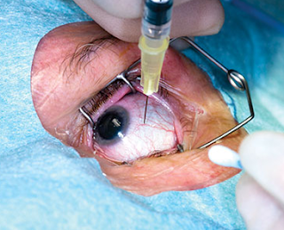
This shocking study has shown that all the millions of dollars poured into injecting the eyeballs of patients who have diabetes and macular edema (a major cause of vision loss in diabetics) with ok vision, does not seem to help save vision compared to 2 years of sole observation. 


This study by the DRCR Retina Network has provided valid level 1 evidence that there is 
no statistical difference in risk of vision loss at 2years and no harm to visual function if a patient decides to WAIT to initiate anti-VEGF therapy until there is “clinically meaningful changes in visual acuity” (meaning visual acuity decreased by 1 eye chart line on 2 consecutive visits or by 2 lines at 1 visit).
Here is what they did:
1. All patients had diabetic macular edema and good visual acuity
2. Each was randomly assigned equally to 3 treatment strategies
1) anti-VEGF therapy: injections into eyeball every 4wks pending OCT/retinal thickness measures and vision
2) intraocular laser of macula
3) observation
3. All were followed up for 2 years. 
What they found:
1. No statistically significant differences among te 3 treatment groups in the primary outcome, which was the proportion of participants who experienced decreased visual acuity of 5 or more letters from baseline.
What this means:
1.  Tight glycemic control (meaning super low-carb/ low sugar diet) and blood pressure control, as well as appropriate treatment of high cholesterol, are super important as it may be equal to the best drugs out there.  **See Amazon video on FASTING & Diet suggestions: controversial to talk to your MD about it (though most MDs do not know much about nutrition as it is not really emphasized in most medical schools). https://www.amazon.com/Science-Fasting-Sylvie-Gilman/dp/B075824XCB/ref=pd_ys_iyr10
2. Patients may not have to risk enophthalmitis with every injection if vision with macular edema is acceptable. 
3. Cost savings: 2011 to 2015, ranibizumab and aflibercept accounted for 12% of the Medicare Part B drug claim spending
Editorial
April 29, 2019

Patients With Good Vision and Diabetic Macular Edema Involving the Center of the MaculaTo Treat or Not to Treat?

JAMA. Published online April 29, 2019. doi:10.1001/jama.2019.5793

Despite the availability of therapies for diabetic retinopathy, it is imperative that clinicians educate patients about the importance of medical therapies including tight glycemic control and blood pressure control as well as appropriate treatment of dyslipidemia in reducing the risk of development and progression of diabetic retinopathy and other microvascular complications.15,16 The volunteers who participated in these clinical trials maintained reasonably good hemoglobin A1C levels. Treating physicians need to consider the generalizability of the study results to their patients and the individual patient’s medical status. These treatment strategies for diabetic macular edema for eyes with good vision also depend on the continued adherence of patients, who have numerous comorbidities, to their follow-up. Treating physicians will need to judiciously determine the best treatment option for their patients with diabetic macular edema and good vision. Once again, the DRCR Retina Network has provided valid level 1 evidence to guide physicians whether (and how) to treat or not to treat persons with diabetic retinopathy, including those with good visual acuity.


Original Investigation
April 29, 2019

Effect of Initial Management With Aflibercept vs Laser Photocoagulation vs Observation on Vision Loss Among Patients With Diabetic Macular Edema Involving the Center of the Macula and Good Visual AcuityA Randomized Clinical Trial

JAMA. Published online April 29, 2019. doi:10.1001/jama.2019.5790
Key Points

Question  For patients with eyes having diabetic macular edema involving the macular center and vision 20/25 or better, what is the effect on vision loss of initial management with aflibercept vs laser photocoagulation vs observation, with aflibercept added to laser photocoagulation and observation if vision worsens?
Findings  In this randomized clinical trial of 702 eyes, a 5-letter or more decrease in visual acuity at 2 years was not significantly different between groups initially managed with aflibercept (16%), laser photocoagulation (17%), and observation (19%).
Meaning  Among eyes with diabetic macular edema involving the macular center and good visual acuity, there was no significant difference in vision loss at 2 years whether eyes were initially managed with aflibercept, laser photocoagulation, or observation.

Abstract
Importance  Intravitreous injections of antivascular endothelial growth factor agents are effective for treating diabetic macular edema (DME) involving the center of the macula (center-involved DME [CI-DME]) with visual acuity impairment (20/32 or worse). The best approach to treating patients with CI-DME and good visual acuity (20/25 or better) is unknown.
Objective  To compare vision loss at 2 years among eyes initially managed with aflibercept, laser photocoagulation, or observation.
Design, Setting, and Participants  Randomized clinical trial conducted at 91 US and Canadian sites among 702 adults with type 1 or type 2 diabetes. Participants had 1 study eye with CI-DME and visual acuity of 20/25 or better. The first participant was randomized on November 8, 2013, and the final date of follow-up was September 11, 2018.
Interventions  Eyes were randomly assigned to 2.0 mg of intravitreous aflibercept (n = 226) as frequently as every 4 weeks, focal/grid laser photocoagulation (n = 240), or observation (n = 236). Aflibercept was required for eyes in the laser photocoagulation or observation groups that had decreased visual acuity from baseline by at least 10 letters (≥ 2 lines on an eye chart) at any visit or by 5 to 9 letters (1-2 lines) at 2 consecutive visits.
Main Outcomes and Measures  The primary outcome was at least a 5-letter visual acuity decrease from baseline at 2 years. Antiplatelet Trialists’ Collaboration adverse events (defined as myocardial infarction, stroke, or vascular or unknown death) were reported.
Results  Among 702 randomized participants (mean age, 59 years; 38% female [n=264]), 625 of 681 (92% excluding deaths) completed the 2-year visit. For eyes with visual acuity that decreased from baseline, aflibercept was initiated in 25% (60/240) and 34% (80/326) in the laser photocoagulation and observation groups, respectively. At 2 years, the percentage of eyes with at least a 5-letter visual acuity decrease was 16% (33/205), 17% (36/212), and 19% (39/208) in the aflibercept, laser photocoagulation, and observation groups, respectively (aflibercept vs laser photocoagulation risk difference, −2% [95% CI, −9% to 5%]; relative risk, 0.88 [95% CI, 0.57-1.35; P = .79]; aflibercept vs observation risk difference, −3% [95% CI, −11% to 4%]; relative risk, 0.83 [95% CI, 0.55-1.27; P = .79]; laser photocoagulation vs observation risk difference, −1% [95% CI, −9% to 6%]; relative risk, 0.95 [95% CI, 0.64-1.41; P = .79]). Antiplatelet Trialists’ Collaboration vascular events occurred in 15 (7%), 13 (5%), and 8 (3%) participants in the aflibercept, laser photocoagulation, and observation groups.
Conclusions and Relevance  Among eyes with CI-DME and good visual acuity, there was no significant difference in vision loss at 2 years whether eyes were initially managed with aflibercept or with laser photocoagulation or observation and given aflibercept only if visual acuity worsened. Observation without treatment unless visual acuity worsens may be a reasonable strategy for CI-DME.
Shopping Cart