I have seen now 8 patients whose meibography looks better after Lipflow.
Meibography does a pretty good job of trying to objectively demonstrate and follow meibomian gland atrophy. There are inherent errors with many imaging technologies, and meibography is not perfect.
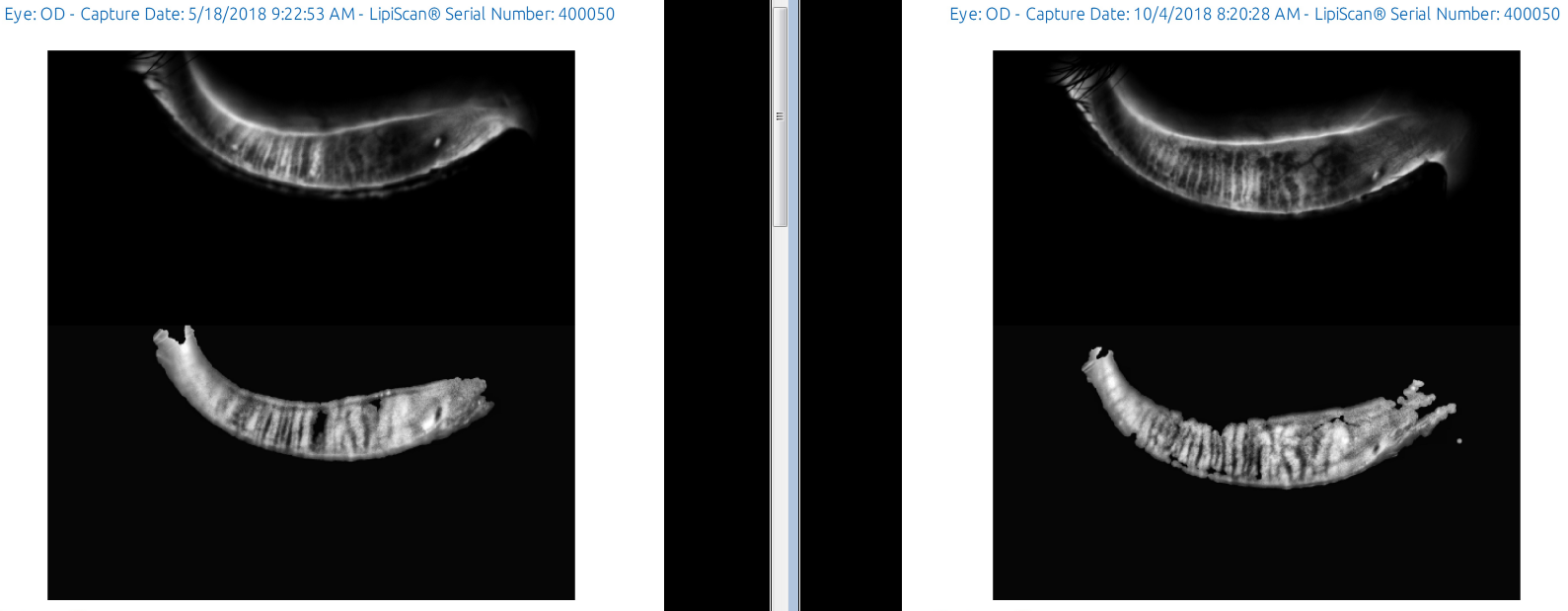
This patient had glands in the area, to begin with so the heat and pulsating action of Lipiflow likely improved oil production.
If a patient has no glands, it is likely too late for Lipiflow to have any effect. I have not seen yet a completely scarred gland return after Lipiflow but I have seen this after PRP insertion after meibomian gland probing, for instance: (update: we presented our research and findings on this at an international conference in Washington DC in 2018.)
This patient had Lipiflow about 4months ago. She had an improvement in her dry eye symptoms but it was not a cure. Her oil expression from the glands that appeared to be atrophied (ie scarred or dysfunctional) was improved.
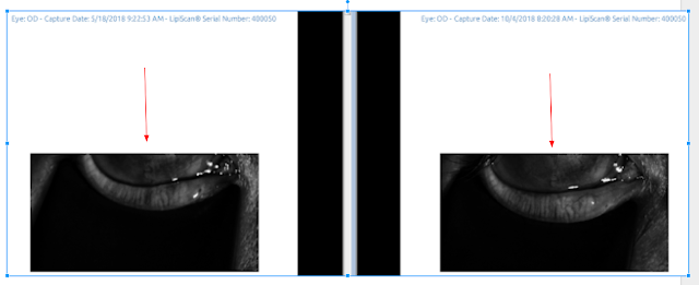
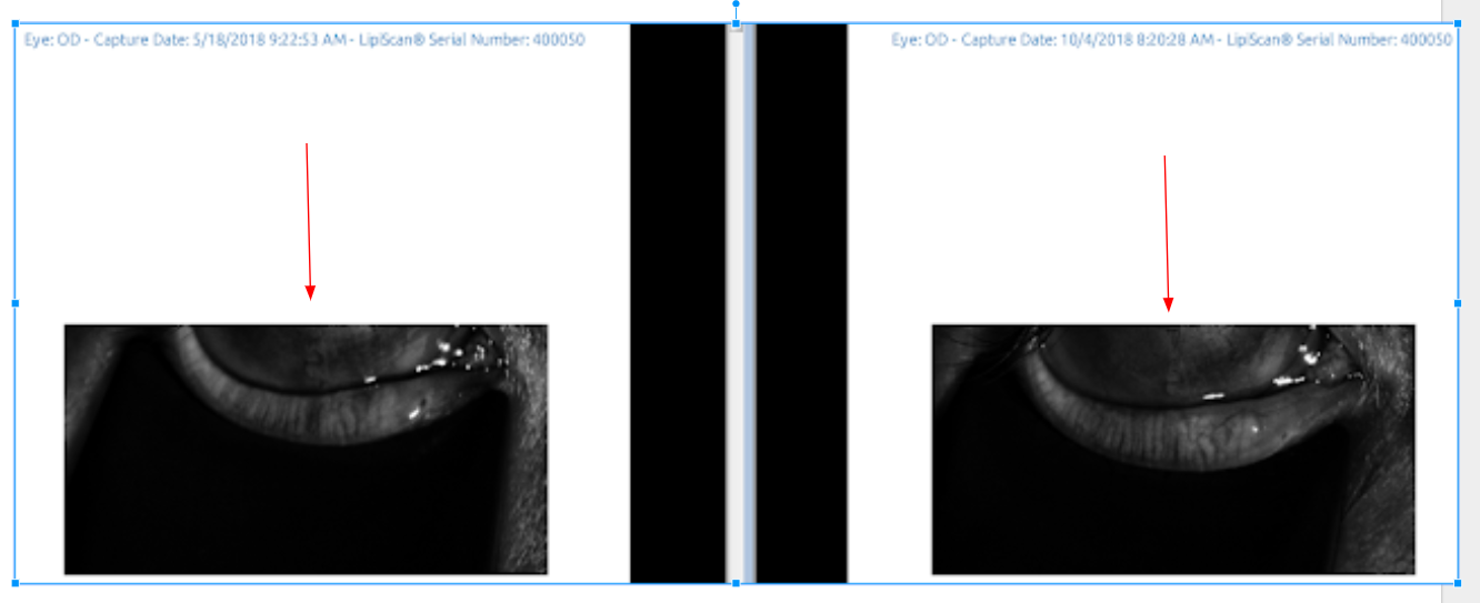
See the dates below and look at the red arrow of the right lower lid. The left lower lid also looks better.
Lipiflow does slow down gland atrophy/loss in an attempt to save the glands, but it is not a cure for dry eyes which is usually due to multiple factors that may not be able to be remedied (ie, genetics, rosacea, aging, hormonal changes).
The only way to 100% prove Lipiflow helped “regrow meibomian glands” in this patient would be to take a sample of the meibomian gland before and after Lipiflow and send to pathology which is not possible yet. So for now, we rely on meibography to see what is going on within the meibomian glands. (My sister at Stanford and I keep talking about meibomian gland transplantation but we are not there yet.)
We are doing Lipiflow on younger and younger patients each year. The youngest we have treated so far is 12 years old because she already has significant corneal scar tissue due to damaged meibomian glands. This is off label by the FDA but I do think it is medically necessary when we see meibomian gland atrophy.
I think the standard of care will be to “medically recommended” Lipiflow if we see even just 1 gland loss as the risk of doing nothing may be devastating with more patients coming in with chronic, debilitating eye pain.
SLC
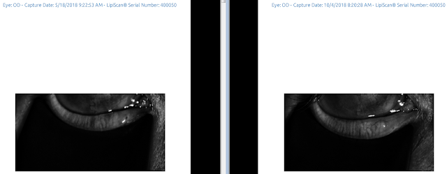
Below is an example of 1 of these patients. There Right EYE (OD) had a stye in the past and the dark line where the red arrow points below on the left, shows where the scar from the stye was located. After the Lipiflow, it appears that the gland has regrown. There was also oil coming out of that gland.
↓