
What is SPK? Superficial Punctate Keratitis
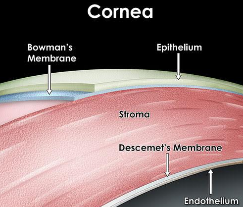
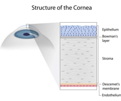
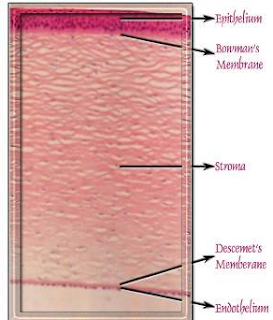
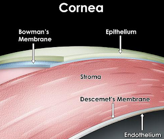
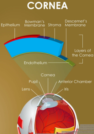 Superficial Punctate Keratitis means that the surface of the Cornea is so dry from Inflammation (-itis…thus keratitis: -kera means Cornea), that the Cornea’s surface epithelial cells have died and left a bare area of underlying Bowman’s membrane (the layer under the epithelium). When we put a yellow drop called Fluoresceine, it stains Bowman membrane cells and not healthy epithelial cells. This is how we can see under the microscope that a patient has dry eyes.
Why do Corneal Epithelial Cells Die?
Corneal Epithelial Cells die from:
1. Direct trauma, like a scratch on your eye or corneal abrasion.
2. Chemicals: getting an acid or base will kill these cells and many other important eye surface cells.
3. LASIK and PRK: we cut the cornea in LASIK or remove the epithelial cells in PRK, and thus purposely remove these cells to sculpt the shape of the cornea to help get a patient glasses free. The risk of doing this is that in some patients, especially if they have decreased Meibomian Glands and/or Dry Eye, the epithelial cells and maybe even the corneal nerves, do not grow back “normally” and the patient feels dry eyes that does not go away.
4. Dry Eye: this is a big issue. Dry Eye can be caused by many things, genetics, aging, hormonal changes, chronic computer use (see below), Meibomian Gland Dysfunction
5. Meibomian Gland Dysfunction: I think a patient can hit a tipping point where the extent of Meibomian Gland loss and oil production loss is significant enough to directly kill corneal epithelial cells. Likely with decreased meibomian glands and oil production, the corneal epithelial cells hit a point chemically where they behave as if to say “I can’t take this milieu/environment any more: I guess I will just disappear.” This then shows us SPK.
We can now image Meibomian Glands clearly for the first time ever. I would venture to say that a patient should not have LASIK or PRK unless they see for themselves their Meibomian Glands on LipiView or LipiScan. This will give them a better idea of what the risk is of having chronic dry eye, chronic halos/glare, chronic discomfort/foreign body sensation, chronic decreased quality of vision even with 20/20 vision which is a real complaint in LASIK and PRK patients.
If the meibomian glands loss is severe, I do not think that patient should have LASIK or PRK. The risk is too great of chronic debilitating eye discomfort, pain, and vision loss.
Normal Meibomian Glands should look like “White Piano Keyes Filled with Oil” On Meibography or LipiScan or LipiView Machines seen on bottom left.
Aging, genetics, chronic blepharitis and meibomian gland dysfunction, poor diet, smoking, inflammatory issues and disease, radiation, chemotherapy, previous surgery, and I must add Chronic computer/video game/cell phone use (ie, Computer Vision Syndrome or a new term I will coin called Decreased Blinking Quality Syndrome or Decreased Blinking Rate Syndrome which can happen to even avid readers, skiers, etc) can destroy these precious glands and cause them to never function again. This loss of meibomian glands, decreases the ability of the tears to lubricate the cornea properly, which then leads to the death of the corneal epithelium, thus giving SPK. The death of the corneal epithelium, though is reversible.
To date, I have had 5 patients whose Meibomian Glands showed “growth” or improvement after LipiFlow or Intense Pulse Light or Meibomian Gland Probing. The current thought, though is that once a meibomian gland is gone, it is gone for good. I hope I am right.
On the right you can see:
Severe Meibomian Gland Loss
